. 2020 Mar 3;117(9):4971-4982.
doi: 10.1073/pnas.1913904117. Epub 2020 Feb 19.

The process of Lewy body formation, rather than simply α-synuclein fibrillization, is one of the major drivers of neurodegeneration

Affiliations

The process of Lewy body formation, rather than simply α-synuclein fibrillization, is one of the major drivers of neurodegeneration

Anne-Laure Mahul-Mellier et al. Proc Natl Acad Sci U S A. .

Abstract

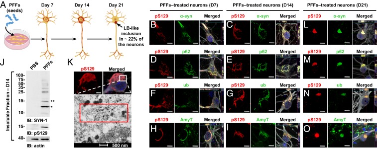
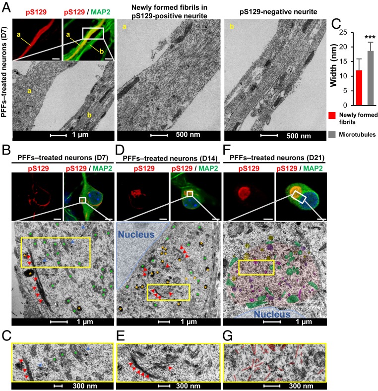
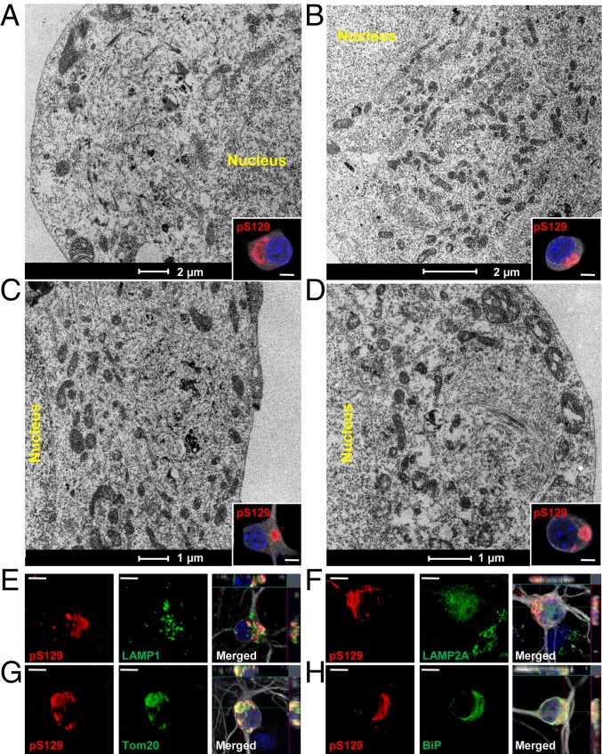
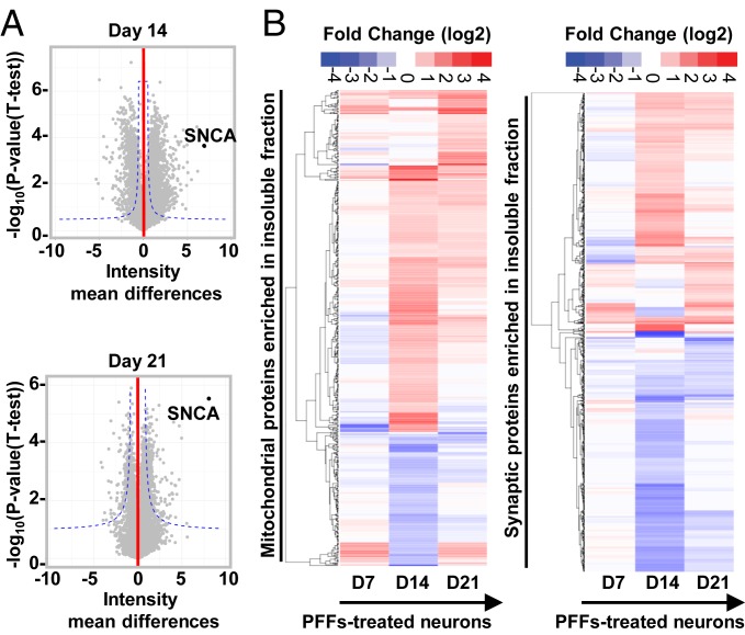
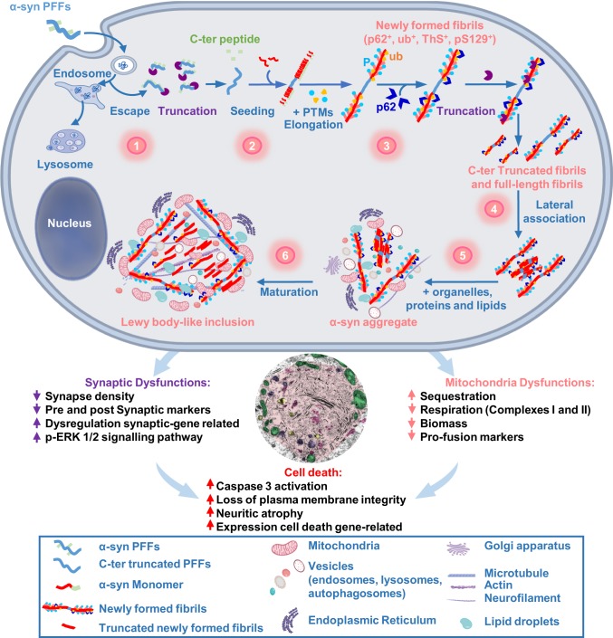
Parkinson's disease (PD) is characterized by the accumulation of misfolded and aggregated α-synuclein (α-syn) into intraneuronal inclusions named Lewy bodies (LBs). Although it is widely believed that α-syn plays a central role in the pathogenesis of PD, the processes that govern α-syn fibrillization and LB formation remain poorly understood. In this work, we sought to dissect the spatiotemporal events involved in the biogenesis of the LBs at the genetic, molecular, biochemical, structural, and cellular levels. Toward this goal, we further developed a seeding-based model of α-syn fibrillization to generate a neuronal model that reproduces the key events leading to LB formation, including seeding, fibrillization, and the formation of inclusions that recapitulate many of the biochemical, structural, and organizational features of bona fide LBs. Using an integrative omics, biochemical and imaging approach, we dissected the molecular events associated with the different stages of LB formation and their contribution to neuronal dysfunction and degeneration. In addition, we demonstrate that LB formation involves a complex interplay between α-syn fibrillization, posttranslational modifications, and interactions between α-syn aggregates and membranous organelles, including mitochondria, the autophagosome, and endolysosome. Finally, we show that the process of LB formation, rather than simply fibril formation, is one of the major drivers of neurodegeneration through disruption of cellular functions and inducing mitochondria damage and deficits, and synaptic dysfunctions. We believe that this model represents a powerful platform to further investigate the mechanisms of LB formation and clearance and to screen and evaluate therapeutics targeting α-syn aggregation and LB formation.

Keywords: Lewy body; Parkinson’s disease; aggregation; seeding; α‐synuclein.


Conflict of interest statement

The authors declare no competing interest.

Figures

Fig. 1.
Fig. 1.
At early stage, α-syn–seeded fibrillar aggregates share the immunohistochemical but not the structural and morphological features of the bona fide human LBs. (A) Seeding model in primary hippocampal neurons. (BI and LO) Temporal analysis of α-syn aggregates formed at D7 (B, D, F, and H), at D14 (C, E, G, and I), and at D21 (LO) in PFF-treated neurons. Aggregates were detected by ICC using either pS129 (81a) in combination with total α-syn (epitope: 1–20) (B, C, and L) or with the Amytracker dye (AmyT; H, I, and O) or pS129 (MJFR13) in combination with p62 (D, E, and M) or ubiquitin (F, G, and N) antibodies. Neurons were counterstained with MAP2 antibody and the nucleus with DAPI staining. (J) Western blot analysis. Total α-syn, pS129, and actin were detected by SYN-1, pS129 (MJFR13), and actin antibodies, respectively. Monomeric α-syn (15 kDa) is indicated by a double asterisk; C-terminal truncated α-syn (12 kDa) is indicated by a single asterisk; the higher molecular weights corresponding to the newly formed fibrils are detected from 25 kDa to the top of the gel. (K) At D14, pS129-positive aggregates imaged by confocal microscopy was then examined by EM after immunogold labeling. (Scale bars: BI and LO, 10 μm; K, Upper, 5 μm.)
Fig. 2.
Fig. 2.
The formation and maturation of LB-like inclusions require the lateral association and the fragmentation of the newly formed α-syn fibrils over time. At the indicated time, PFF-treated neurons were fixed, ICC against pS129-α-syn was performed and imaged by confocal microscopy (A, B, D, and F, Top), and the selected neurons were then examined by EM (A, B, D, and F, Bottom). (A, a) Neurite with pS129-positive newly formed α-syn fibrils. (A, b) A neurite negative for pS129 staining. (A, c) Graph representing the mean ± SD of the width of the microtubules compared to the newly formed fibrils at D7. **P < 0.001 (Student’s t test for unpaired data with equal variance), indicating that this parameter can be used to discriminate the newly formed fibrils from the cytoskeletal proteins. (Scale bars: A, B, D, and F, Top, 10 μm.) (CG) Representative images at higher magnifications corresponding to the area indicated by a yellow rectangle in EM images shown in B, D, and F.
Fig. 3.
Fig. 3.
Maturation of α-syn–seeded aggregates into LB-like inclusions is accompanied by the sequestration of organelles and the endomembrane system. (AD) At D21, PFF-treated neurons were fixed, ICC against pS129-α-syn was performed and imaged by confocal microscopy (Insets) and selected neurons were examined by EM. Representative images of LB-like inclusions exhibiting a ribbon-like morphology (A and B) or compact and rounded structures (C and D). Mitochondria, autophagosomes, endosome, and lysosome-like vesicles were detected inside or at the edge of these inclusions (see also Fig. S6 AE). (EH) LB-like inclusions were stained by pS129 in combination with organelles markers LAMP1 (late endosomes) (E), LAMP2A (autophagosomes) (F), Tom20 (mitochondria) (G), or BiP (ER) (H). Neurons were counterstained with MAP2 antibody and nucleus with DAPI (see also Fig. S6 FI). (Scale bars: AD, Insets, 5 μm; EH, 10 μm.)
Fig. 4.
Fig. 4.
Temporal proteomic analyses of the protein contents found in the insoluble fraction of the PFF-treated neurons reveals a high increase in proteins related to the endomembrane system. (A) Insoluble proteins from neurons treated with PBS and PFFs for 7, 14, and 21 d were extracted and analyzed using LC-MS/MS. Identified proteins were plotted using volcano plot. Dotted lines represent the false discovery rate < 0.05 and threshold of significance SO = 1 assigned for the subsequent analysis. A detailed list of the hits is available in Dataset S1. Classifications of the proteins significantly enriched in the insoluble fractions of the PFF-treated neurons at D14 and D21 by cellular component and biological processes using gene ontology (GO) and DAVID enrichment analyses are shown in SI Appendix, Fig. S8 B and C. (B) Heat maps representing color-coded fold-change levels of mitochondrial (Left) and synaptic (Right) proteins present in the insoluble fraction in PFF-treated neurons.
Fig. 5.
Fig. 5.
Gene-expression level changes during the formation of the newly formed fibrils and their maturation into LB-like inclusions. Temporal transcriptomic analysis of the gene-expression level in PBS-treated neurons vs. PFF-treated neurons treated for 7, 14, or 21 d. Genes with an absolute log2 fold change greater than 1 and a false discovery rate less than 0.01 were considered as significantly differentially expressed (SI Appendix, Fig. S9). Dataset S2 depicts the number of genes up- or down-regulated in PFF-treated neurons over time. (A and B) Log2 fold changes of the mitochondrial genes (A and Dataset S3), and the synaptic genes (B and Dataset S4) expression levels are represented over time. “*” indicates a significant up- or down-regulation in the gene-expression level.
Fig. 6.
Fig. 6.
Dynamics of Lewy body formation and maturation induce mitochondrial alterations. (A) High-resolution respirometry measurements. Statistical results were obtained from two-way repeated measurement ANOVAs based on a minimum of four independent experiments. (B and C) Proteomic data on proteins implicated in mitochondrial dysfunction. (D) Mitochondrial membrane potential was assessed fluorometrically from cells loaded with TMRE. (E) Measurement of mitotracker green level, which labels mitochondrial proteins independently of mitochondrial membrane potential. (F) Western blot analyses of total fractions immunoblotted with antibodies against components of the OXPHOS complexes I-V, mitofusin 2, OPA1, citrate synthase, and VDAC1 proteins. Actin was used as the loading control. The graphs (DF), represent the mean ± SD of a minimum of three independent experiments. *P < 0.05, **P < 0.005, ***P < 0.0005 (ANOVA followed by Tukey honest significant difference [HSD] post hoc test, PBS- vs. PFF-treated neurons). #P < 0.05, ###P < 0.0005 (D14 vs. D21 PFF-treated neurons). a.u., arbitrary unit.
Fig. 7.
Fig. 7.
Synaptic dysfunctions were associated with the formation and maturation of the LB-like inclusions. (A) The levels of Synapsin I, PSD95, and ERK 1/2 were assessed by Western blot over time. Actin was used as the loading control. The graphs represent the mean ± SD of a minimum of three independent experiments. (B and C) Synaptic area decreases in PFF-treated neurons from D14. (B) Aggregates were detected by ICC using pS129 (MJFR13) and Synapsin I antibodies. Neurons were counterstained with MAP2 antibody, and the nucleus with DAPI. (Scale bars: 5 μm.) (C) Measurement of the synaptic area was performed over time. (A and C) ANOVA followed by Tukey honest significant difference [HSD] post hoc test was performed. *P < 0.05, **P < 0.005 (PBS- vs. PFF-treated neurons). ##P < 0.005 (D14 vs. D21 PFF-treated neurons). a.u., arbitrary unit.
Fig. 8.
Fig. 8.
The dynamics of Lewy body formation, rather than simply α-syn fibrillization, is the primary cause of mitochondrial alterations, synaptic dysfunction, and neurodegeneration. Formation of α-syn inclusions in the context of the neuronal seeding requires a sequence of events starting with 1) the internalization and cleavage of PFF seeds (D0–D1); followed by 2) the initiation of the seeding by the recruitment of endogenous α-syn (D0–D4); 3) fibril elongation along with the incorporation of posttranslational modifications (PTMs), such as phosphorylation at residue S129 and ubiquitination (D4–D14); 4) formation of seeded filamentous aggregates (D7) that are fragmented and laterally associated over time (D7–D21); and 5–6) the packing of the fibrils into LB-like inclusions (D14–D21), which have a morphology similar to the bona fide LB detected in human synucleinopathies (D21), is accompanied by the sequestration of organelles, endomembranes, proteins, and lipids. Our integrated approach using advanced techniques in EM, proteomics, transcriptomics, and biochemistry clearly demonstrate that LB formation and maturation impaired the normal functions and biological processes in PFF-treated neurons, causing mitochondrial alterations and synaptic dysfunctions that result in a progressive neurodegeneration.

References

    1. Peng C., Gathagan R. J., Lee V. M., Distinct α-synuclein strains and implications for heterogeneity among α-synucleinopathies. Neurobiol. Dis. 109, 209–218 (2018). - PMC - PubMed
    1. Peelaerts W., Bousset L., Baekelandt V., Melki R., ɑ-Synuclein strains and seeding in Parkinson’s disease, incidental Lewy body disease, dementia with Lewy bodies and multiple system atrophy: Similarities and differences. Cell Tissue Res. 373, 195–212 (2018). - PubMed
    1. Forno L. S., Concentric hyalin intraneuronal inclusions of Lewy type in the brains of elderly persons (50 incidental cases): Relationship to parkinsonism. J. Am. Geriatr. Soc. 17, 557–575 (1969). - PubMed
    1. Forno L. S., DeLanney L. E., Irwin I., Langston J. W., Electron microscopy of Lewy bodies in the amygdala-parahippocampal region. Comparison with inclusion bodies in the MPTP-treated squirrel monkey. Adv. Neurol. 69, 217–228 (1996). - PubMed
    1. Forno L. S., Norville R. L., Ultrastructure of Lewy bodies in the stellate ganglion. Acta Neuropathol. 34, 183–197 (1976). - PubMed

Publication types

Substances