. 2020 Feb 19;12(531):eaay4006.
doi: 10.1126/scitranslmed.aay4006.

A geometrically adaptable heart valve replacement

Affiliations

A geometrically adaptable heart valve replacement

Sophie C Hofferberth et al. Sci Transl Med. .

Abstract

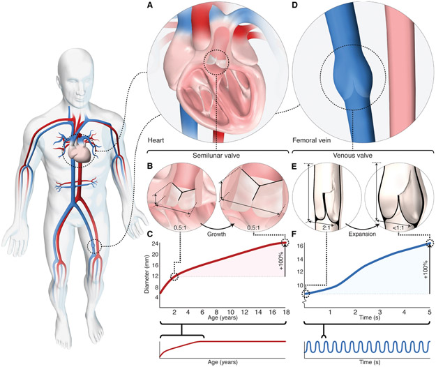
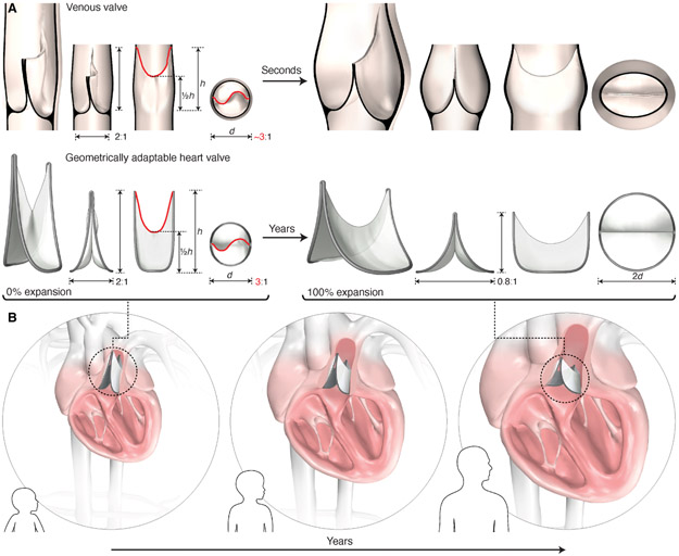
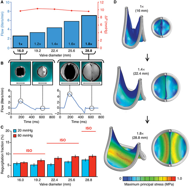
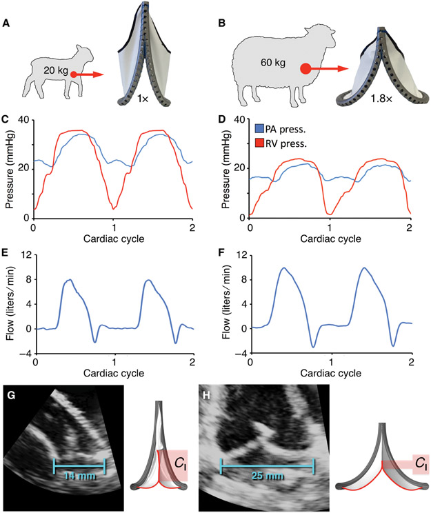
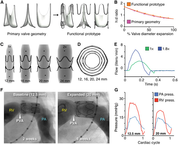
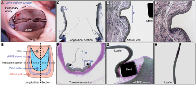
Congenital heart valve disease has life-threatening consequences that warrant early valve replacement; however, the development of a growth-accommodating prosthetic valve has remained elusive. Thousands of children continue to face multiple high-risk open-heart operations to replace valves that they have outgrown. Here, we demonstrate a biomimetic prosthetic valve that is geometrically adaptable to accommodate somatic growth and structural asymmetries within the heart. Inspired by the human venous valve, whose geometry is optimized to preserve functionality across a wide range of constantly varying volume loads and diameters, our balloon-expandable synthetic bileaflet valve analog exhibits similar adaptability to dimensional and shape changes. Benchtop and acute in vivo experiments validated design functionality, and in vivo survival studies in growing sheep demonstrated that mechanical valve expansion accommodated growth. As illustrated in this work, dynamic size adaptability with preservation of unidirectional flow in prosthetic valves thus offers a paradigm shift in the treatment of heart valve disease.


Figures

Fig. 1.
Fig. 1.. Geometry and function of human arterial and venous valves.
(A) Schematic depicting arterial semilunar (pulmonary) valve located within the heart. (B) Schematic depicting axial height of human semilunar valve relative to annulus diameter. Axial height is ~1/2 the annulus diameter and remains constant with growth. (C) Graph of semilunar valve (pulmonary) annulus dimensional change with somatic growth; ~2-fold increase in diameter is observed between the ages of 2 to 18 years in healthy children (graph adapted from reference 23). (D) Schematic depicting native venous valve located within vein of lower extremity. (E) Schematic depicting leaflet dimensions of human venous valve, at rest (left) and during expansion (right). Axial height is ~2 times the vessel diameter and leaflet contact length (coaptation) is up to half the vessel diameter (30, 31). As the vein expands radially with increased volume load, the venous valve aspect ratio decreases (<1:1) and the sinuses dilate to maintain leaflet contact and ensure unidirectional blood flow. (F) Graph of vein diameter in response to large changes in blood volume occurring over a period of seconds. Up to a 4-fold change in vein diameter occurs over a period of seconds (33).
Fig. 2.
Fig. 2.. Design of a biomimetic geometrically adaptable heart valve.
(A) Biomimetic valve design inspired by the geometric profile of the human venous valve. Design is defined by leaflet dimensions, leaflet attachment geometry, and valve expansion geometry. Key leaflet dimensions include the baseline aspect ratio (2:1), leaflet mid-height (1/2h), and free-edge length (red line). The leaflet attachment geometry replicates the 3-dimensional geometric curve created by the venous valve leaflet attachment to the vein wall. The leaflet attachment perimeter is fixed in length, therefore radial expansion is accompanied by a reduction in valve height (aspect ratio decreases to 0.8:1 at 2X expansion; expansion shown on right). (B) Schematic representation of the dynamic biomimetic valve geometry as it adapts to the increasing heart dimensions of a growing child via periodic mechanical balloon expansion.
Fig. 3.
Fig. 3.. Flow loop testing and computational modeling of the biomimetic bileaflet valve.
(A-C) Valve prototypes tested at different states of diametric expansion (1X to 1.8X) in an in vitro circulatory flow loop, under physiologic left and right heart conditions. (A) Transvalvular (ΔP) pressure gradient (red line) at each stage of valve expansion (1X to 1.8X). Flow adjusted to match valve size and growing child physiology. (B) Top view photographs of 1X (black arrow measures 16mm) and 1.8X (black arrow measures 28.8mm) expanded valves within in vitro flow loop system, with leaflets in open (left) and closed (right) position, with their corresponding plots demonstrating flow profiles of the 1X and 1.8X expanded valve geometries. (C) Leaflet coaptation (closing function) under right (20 mmHg, blue bars) and left (80 mmHg, red bars) heart loads. Error bars represent ± 1 standard deviation. Red horizontal lines indicate the upper bound industry standard (International Standards Organization; ISO 5840-2:2015) for prosthetic valve regurgitation % under left heart loads. (D) Finite element model of leaflet stresses in quasi-static loaded state at 3 different states of valve expansion (1X:16mm ID, 1.4X: 22.4mm ID, 1.8X: 28.8mm ID). Leaflet material is 0.1 mm thickness ePTFE, and the results shown are simulated under right heart (20 mmHg) loads. All plots presented are based on the same scale of maximum principal stress, ranging between 0 to 1 MPa. ID = Internal diameter
Fig. 4.
Fig. 4.. Acute in vivo validation of primary biomimetic valve function in juvenile and adult sheep.
(A and B) Fabricated prototypes implanted at two polar expansion states (1X and 1.8X) in the native pulmonary valve position of juvenile (N = 4) and adult (N = 4) sheep. Representative plots showing right ventricular and pulmonary artery pressures for the (C) 1X and (D) 1.8X valve geometries. Corresponding pulmonary artery flow for the (E) 1X and (F) 1.8X valve geometries. Representative echocardiographic images of implanted valves, demonstrating change in coaptation length (Cl) between the (G) baseline (1X, 14 mm ID) and (H) fully expanded (1.8X, 25 mm ID) geometries. ID = internal diameter.
Fig. 5.
Fig. 5.. Functional prototype design and in vivo proof-of-concept of valve expansion in a growing lamb model.
(A) CAD models of primary valve geometry and functional prototype containing added structural support features to optimize valve geometry for mechanical expansion. (B) Valve expansion geometry (change in aspect ratio with radial expansion) for the primary valve geometry (pink line) and functional prototype (orange line), h:d = height to diameter ratio (C) X-ray images of laser-cut stainless-steel functional valve prototypes being expanded via serial balloon dilation. Black dots are x-ray opaque markers on the balloon, denoting zone of constant diameter (D) Top-view x-ray transmission images showing geometry of the functional prototype with expansion from 1X to 2X diameter. (E) In vitro flow loop testing of functional prototype at two polar expansion states, 1X (green) and 1.8X (blue). Plots show the valve flow cycle at both expansion states. (F) Representative right ventricular (RV) angiograms in a lamb showing the expandable biomimetic valve prototype in its baseline (12.5 mm ID) and expanded (20 mm ID, after 3 separate balloon dilation procedures) configuration at 2- and 9-weeks post implantation, respectively. RV = right ventricle, PA = pulmonary artery, PVA = native pulmonary valve annulus (G) Representative right ventricular and pulmonary artery pressures recorded at two states of valve expansion, (left:1X, 12.5mm ID) and (right: 1.6X, 20mm ID) at 2 and 9 weeks after implantation.
Fig. 6.
Fig. 6.. Macroscopic and histological analysis of biomimetic valve prototype implanted for 10 weeks in a growing lamb.
(A) Representative photograph of valve outflow surface at time of valve explant. White dotted arrow indicates valve internal diameter (20mm). (B) Schematic diagram demonstrating method of device sectioning after performing plastic embedding of the explanted specimen (valve and attached pulmonary artery). (C) Representative elastin stained longitudinal section of explanted valve specimen (image width = 30 mm). Boxed region shown at higher magnification in (D). (D) Higher magnification of native pulmonary artery wall, demonstrating tissue architecture at the site of vessel-stent contact and region of artery wall proximal and distal to vessel-stent interface (image width = 2.5 mm). Boxed region shown at higher magnification in (E). (E) Higher magnification of native pulmonary artery wall at site of vessel-stent contact (image width = 1 mm). (F) Representative Hematoxylin and Eosin stained transverse section of explanted valve (image width = 20 mm). Boxed regions shown at higher magnification in (G) and (H). (G) Higher magnification image of valve leaflet at the site of stent attachment with adjacent ePTFE sleeve (image width = 2.5 mm). (H) Higher magnification image showing the mid-section of the valve leaflet (image width = 2.5 mm). All macroscopic and histological images shown are from the same animal. A total of 7 animals received valve implants (See fig. S16, fig. S17).

References

    1. van der Linde D, Konings EE, Slager MA, Witsenburg M, Helbing WA, Takkenberg JJ, Roos-Hesselink JW, Birth prevalence of congenital heart disease worldwide: a systematic review and meta-analysis. J Am Coll Cardiol 58, 2241–2247 (2011). - PubMed
    1. Bernier PL, Stefanescu A, Samoukovic G, Tchervenkov CI, The challenge of congenital heart disease worldwide: epidemiologic and demographic facts. Semin Thorac Cardiovasc Surg Pediatr Card Surg Annu 13, 26–34 (2010). - PubMed
    1. Gilboa SM, Devine OJ, Kucik JE, Oster ME, Riehle-Colarusso T, Nembhard WN, Xu P, Correa A, Jenkins K, Marelli AJ, Congenital Heart Defects in the United States: Estimating the Magnitude of the Affected Population in 2010. Circulation 134, 101–109 (2016). - PMC - PubMed
    1. Benjamin EJ, Blaha MJ, Chiuve SE, Cushman M, Das SR, Deo R, de Ferranti SD, Floyd J, Fornage M, Gillespie C, Isasi CR, Jimenez MC, Jordan LC, Judd SE, Lackland D, Lichtman JH, Lisabeth L, Liu S, Longenecker CT, Mackey RH, Matsushita K, Mozaffarian D, Mussolino ME, Nasir K, Neumar RW, Palaniappan L, Pandey DK, Thiagarajan RR, Reeves MJ, Ritchey M, Rodriguez CJ, Roth GA, Rosamond WD, Sasson C, Towfighi A, Tsao CW, Turner MB, Virani SS, Voeks JH, Willey JZ, Wilkins JT, Wu JH, Alger HM, Wong SS, Muntner P, American C Heart Association Statistics, S. Stroke Statistics, Heart Disease and Stroke Statistics-2017 Update: A Report From the American Heart Association. Circulation 135, e146–e603 (2017). - PMC - PubMed
    1. Hoffman J, The global burden of congenital heart disease. Cardiovasc J Afr 24, 141–145 (2013). - PMC - PubMed

Publication types

LinkOut - more resources