. 2019 Jun 19;44(1):16-23.
doi: 10.1080/01658107.2019.1615960. eCollection 2020 Feb.

Selective Colour Vision Deficits in Multiple Sclerosis at Different Temporal Stages

Affiliations

Selective Colour Vision Deficits in Multiple Sclerosis at Different Temporal Stages

Neda Anssari et al. Neuroophthalmology. .

Abstract

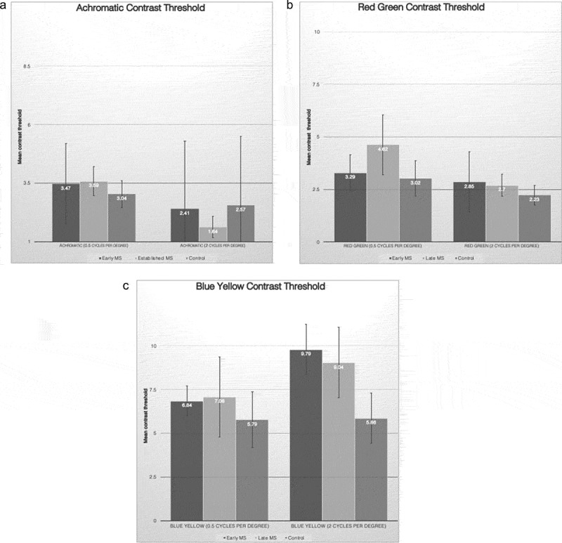
Multiple sclerosis (MS) without optic neuritis causes color-vision deficit but the evidence for selective color deficits in parvocellular-Red/Green (PC-RG) and koniocellular-Blue/Yellow (KC-BY) pathways is inconclusive. We investigated selective color-vision deficits at different MS stages. Thirty-one MS and twenty normal participants were tested for achromatic, red-green and blue-yellow sinewave-gratings (0.5 and 2 cycles-per-degree (cpd)) contrast orientation discrimination threshold. Red-green mean threshold at 0.5cpd in established-MS and blue-yellow mean threshold in all MS participants were abnormal. These findings show blue-yellow versus red-green color test is useful in differentiating MS chronicity, which helps to better understand the mechanism of colour-vision involvement in MS.

Keywords: Multiple sclerosis; blue/yellow; colour vision; psychophysics; red/green.


Figures

Figure 1.
Figure 1.
Horizontal achromatic, vertical red–green and horizontal blue–yellow sinusoidal Gabors presented to participants and controls.
Figure 2.
Figure 2.
(a) Mean contrast threshold for achromatic stimuli at 0.5 and 2 cycles per degree SF. The difference in the mean contrast threshold of early MS, established MS and controls was not statistically significant. Error bars represent ± 0.5 standard deviation. (b) Mean contrast threshold for red–green stimuli at 0.5 and 2 cycles per degree SF. The mean contrast threshold was significantly elevated in established MS at SF of 0.5 compared with controls. Error bars represent ± 0.5 standard deviation. (c) Mean contrast threshold for blue–yellow stimuli at 0.5 and 2 cycles per degree SF. The mean contrast threshold at 2 cycles per degree was significantly higher in early and established MS versus controls. Error bars represent ± 0.5 standard deviation.

References

    1. Harrison AC, Becker WJ, Stell WK.. Colour vision abnormalities in multiple sclerosis. Can J Neurol Sci. 1987;14:279–285. - PubMed
    1. Moura AL, Teixeira RA, Oiwa NN, et al. Chromatic discrimination losses in multiple sclerosis patients with and without optic neuritis using the Cambridge colour test. Vis Neurosci. 2008;25:463–468. doi:10.1017/S0952523808080437. - DOI - PubMed
    1. Gundogan FC, Tas A, Altun S, Oz O, Erdem U, Sobaci G. Colour vision versus pattern visual evoked potentials in the assessment of subclinical optic pathway involvement in multiple sclerosis. Indian J Ophthalmol. 2013;61:100–103. doi:10.4103/0301-4738.99842. - DOI - PMC - PubMed
    1. Al-Hashmi AM, Kramer DJ, Mullen KT. Human vision with a lesion of the parvocellular pathway: an optic neuritis model for selective contrast sensitivity deficits with severe loss of midget ganglion cell function. Exp Brain Res. 2011;215:293–305. doi:10.1007/s00221-011-2896-4. - DOI - PubMed
    1. Travis D, Thompson P. Spatiotemporal contrast sensitivity and colour vision in multiple sclerosis. Brain. 1989;112(Pt 2):283–303. - PubMed

LinkOut - more resources