Case Reports
. 2018 Nov 13;44(1):38-40.
doi: 10.1080/01658107.2018.1542008. eCollection 2020 Feb.

Encephalomyelitis with Retinopathy in Common Variable Immunodeficiency (CVID)

Affiliations
Case Reports

Encephalomyelitis with Retinopathy in Common Variable Immunodeficiency (CVID)

Samuel E Shribman et al. Neuroophthalmology. .

Abstract

Common variable immunodeficiency is the most common primary immunodeficiency and rarely causes neurological manifestations since the introduction of IVIg, but here, the authors present a case of a 31-year-old Afro-Caribbean man who after short non-adherence to his immunoglobulins, develops encephalomyelitis with retinopathy. To the authors' knowledge, this is the first case presented with retinal photographs, OCT, CT, MRI and brain biopsies.

Keywords: Encephalomyelitis; MRI; case report; immunodeficiency; ophthalmology.


Figures

Figure 1.
Figure 1.
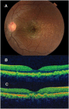
(A) Retinal photograph of the left fundus showing drusen-like abnormalities in the macula. (B) Ocular coherence tomography of the left eye macula showing drusen-like deposits just below the level of the retinal pigment epithelium. (C) Ocular coherence tomography of the left eye macula 12 months later showing persisting drusen-like deposits.
Figure 2.
Figure 2.
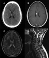
(A) Unenhanced axial CT brain image showing a hyperdense mass within the right callosal body. (B) Axial T1-weighted MRI brain post-gadolinium demonstrating a contrast enhancing mass within right callosal body. (C) Axial T2-weighted MRI brain image showing bilateral, confluent hyper-intensity through the periventricular and deep cerebral white matter. (D) T2-weighted sagittal MR image showing diffuse intramedullary signal change within the cervical cord.
Figure 3.
Figure 3.
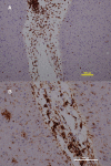
Right frontal lobe biopsy. The white matter shows dilated Virchow-Robin spaces with mild perivascular infiltration by lymphocytes and macrophages. An occasional microglial nodule and scattered apoptosis is seen. There is mildly increasing glial cellularity and pleomorphism consistent with reactive gliosis. Adjacent cortex shows mild gliosis and an occasional perivascular lymphocyte. (A) Immunostaining for CD3 shows moderate numbers of perivascular T-cells and scattered parenchymal T-cells. Immunostaining for CD20 shows no B-cells. (B) Immunostaining for CD68 shows moderate numbers of perivascular macrophages and moderate parenchymal microglial activation.

References

    1. Rudge P, Webster AD, Revesz T, Warner T, Espanol T, Cunningham-Rundles C, Hyman N. Encephalomyelitis in primary hypogammaglobulinaemia. Brain. 1996;119:1–15. - PubMed
    1. Resnick ES, Moshier EL, Godbold JH, Cunningham-Rundles C.. Morbidity and mortality in common variable immune deficiency over 4 decades. Blood. 2012;119(7):1650–1657. doi:10.1182/blood-2011-09-377945. - DOI - PMC - PubMed
    1. Nguyen JT, Green A, Wilson MR, DeRisi JL, Gundling K. Neurologic complications of common variable immunodeficiency: A case report and review of the literature. J Clin Immunol. 2016;36(8):793–800. doi:10.1007/s10875-016-0336-8. - DOI - PubMed
    1. Ziegner UH, Kobayashi RH, Cunningham-Rundles C, Español T, Fasth A, Huttenlocher A, Krogstad P, Marthinsen L, Notarangelo LD, Pasic S, Rieger CH, Rudge P, Sankar R, Shigeoka AO, Stiehm ER, Sullivan KE, Webster AD, Ochs HD. Progressive neurodegeneration in patients with primary immunodeficiency disease on IVIG treatment. Clin Immunol. 2002;102(1):19–24. doi:10.1006/clim.2001.5140. - DOI - PubMed
    1. Fang B, McKeon A, Hinson SR, Kryzer TJ, Pittock SJ, Aksamit AJ, Lennon VA. Autoimmune glial fibrillary acidic protein astrocytopathy: A novel meningoencephalomyelitis. JAMA Neurol. 2016;73(11):1297–1307. doi:10.1001/jamaneurol.2016.2549. - DOI - PubMed

Publication types

LinkOut - more resources