. 2020 Jan 28:2020:5863178.
doi: 10.1155/2020/5863178. eCollection 2020.

ZNF143 Suppresses Cell Apoptosis and Promotes Proliferation in Gastric Cancer via ROS/p53 Axis

Affiliations

ZNF143 Suppresses Cell Apoptosis and Promotes Proliferation in Gastric Cancer via ROS/p53 Axis

Yi Zhang et al. Dis Markers. .

Abstract

Aim: This study was aimed at identifying the role of zinc finger protein 143 (ZNF143) in gastric cancer (GC) progression.

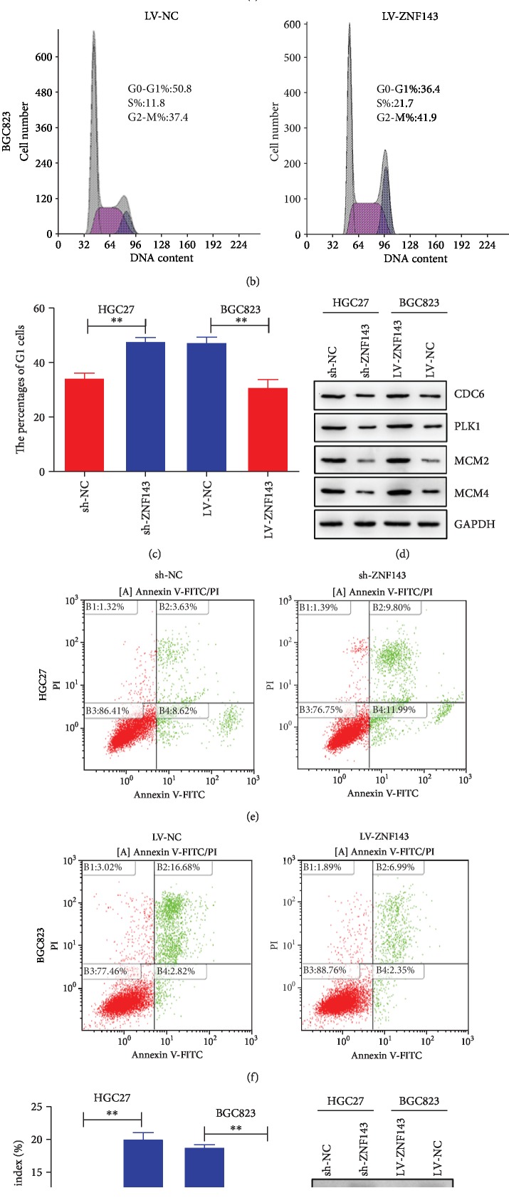
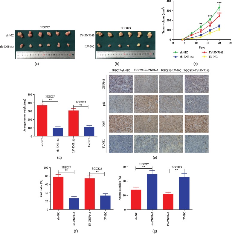
Methods: The impact of ZNF143 on the proliferation ability and apoptosis of GC cells was detected. The expression of ZNF143 and related targeted genes was determined using Western blot analysis. The reactive oxygen species (ROS) level of GC cells was examined using the ROS generation assay. The role of ZNF143 in the proliferation of GC cells in vivo was examined using tumor xenograft assay.

Results: The ectopic overexpression of ZNF143 promoted the proliferation of GC cells, while its knockdown reduced the effect in vitro. The downregulation of ZNF143 facilitated cell apoptosis. ZNF143 decreased the ROS level in GC cells, resulting in the reduction of cell apoptosis. Transfection with p53 reversed the antiapoptotic effect of ZNF143, while pifithrin-α, a specific inhibitor of p53, reduced the apoptosis in ZNF143-knockdown GC cells. However, p53 had no influence on the ROS level in GC cells. p53 played a key role in inhibiting ROS generation in GC cells, thereby inhibiting apoptosis. The transplanted tumor weight and volume were higher in the ZNF143-overexpressed group than in the ZNF143-knockdown group in vivo was examined using tumor xenograft assay.

Conclusion: ZNF143, as a tumor oncogene, promoted the proliferation of GC cells both in vitro and in vivo, indicating that ZNF143 might function as a novel target for GC therapy.in vitro. The downregulation of ZNF143 facilitated cell apoptosis. ZNF143 decreased the ROS level in GC cells, resulting in the reduction of cell apoptosis. Transfection with p53 reversed the antiapoptotic effect of ZNF143, while pifithrin-in vivo was examined using tumor xenograft assay.


Conflict of interest statement

The authors declare no conflicts of interest.

Figures

Figure 1
Figure 1
(a) The expression patterns of GC tumors (n = 408) and normal GC tissues (n = 211) based on The Cancer Genome Atlas (TCGA) and the Genotype-Tissue Expression (GTEx) data in the GEPIA database (http://gepia2.cancer-pku.cn/#analysis). (b) The expression of ZNF143 in GC tumors and corresponding normal tissues using immunohistochemical staining. (c, d) Expression of ZNF143 in HGC27 cells transfected with sh-ZNF143 and in BGC823 cells transfected with LV-ZNF143 lentivirus. (c) The expression of ZNF143 in HGC27 and BGC823 cells analyzed using Western blot analysis. (d) Expression of ZNF143 detected by real-time PCR in HGC27 and BGC823 cells. (e, f) Expression of ZNF143 in HGC27 and BGC823 cells examined using immunofluorescence. P < 0.05, ∗∗P < 0.01, and ∗∗∗P < 0.001. The data were expressed as mean ± standard deviation. GC: gastric cancer; ZNF143: zinc finger protein 143; GAPDH: glyceraldehyde-3-phosphate dehydrogenase; DAPI: 4′,6-diamidino-2-phenylindole dihydrochloride.
Figure 2
Figure 2
ZNF143 promoted the proliferation of GC cells in vitro. (a, b) Effects of ZNF143 on the proliferation ability of GC cells explored using the CCK-8 assay. (c) ZNF143 knockdown inhibited the proliferation of HGC27 cells, and overexpression of ZNF143 promoted the proliferation of BGC823 cells, as detected using EDU assays. (d) Rate of EDU-positive cells. (e) Colony formation assays showing that ZNF143 knockdown inhibited the proliferation of HGC27 cells, while the overexpression of ZNF143 promoted the proliferation of BGC823 cells. (f) Number of clones formed. P < 0.05, ∗∗P < 0.01, and ∗∗∗P < 0.001. The data were expressed as mean ± standard deviation. CCK8: Cell Counting Kit-8; EDU: 5-ethynyl-2′-deoxyuridine.
Figure 3
Figure 3
ZNF143 inhibited cell cycle arrest in the G1 phase and the apoptosis of GC cells. (a, b) Cell cycle distribution of GC cells measured using flow cytometry. (c) Percentage of G1 phase. (d) Protein expression of CDC6, PLK1, MCM2, and MCM4 detected using Western blot analysis. (e, f) Apoptotic rate of GC cells measured using flow cytometry. (g) Apoptotic rate of GC cells. (h) Protein expression levels of p53, Bcl-2, Bax-cl, and Bax detected using Western blot analysis. P < 0.05, ∗∗P < 0.01, and ∗∗∗P < 0.001. The data were expressed as mean ± standard deviation. CDC6: cell division cycle 6; PLK1: polo-like kinase 1; MCM2: minichromosome maintenance complex component 2; MCM4: minichromosome maintenance complex component 4; p53: tumor protein 53; Bcl-2: B-cell lymphoma-2; Bcl-xl: BCL2-like 1; Bax: BCL2 associated X; GAPDH: glyceraldehyde-3-phosphate dehydrogenase.
Figure 4
Figure 4
Overexpression of ZNF143 inhibited p53-dependent ROS-mediated apoptosis in BGC823. (a) Protein level of ZNF143 detected using Western blot assay. (b) Apoptotic rate of GC cells measured using flow cytometry. (c, d) ROS level of GC cells detected using confocal microscopy. (e) Expression of ZNF143 and p53 in GC cells analyzed using Western blot assay. (f) Apoptotic rate of GC cells measured using flow cytometry. (g, h) ROS level of GC cells after different treatments were detected using confocal microscopy. P < 0.05, ∗∗P < 0.01, and ∗∗∗P < 0.001. The data were expressed as mean ± standard deviation. NAC: N-acetylcysteine; ROS, reactive oxygen species.
Figure 5
Figure 5
Knockdown of ZNF143 could promote p53-dependent ROS-mediated apoptosis in HGC27. (a) Protein level of ZNF143 detected using Western blot analysis. (b) Apoptotic rate of GC cells measured using flow cytometry. (c, d) ROS level of GC cells detected using confocal microscopy. (e) Expression of ZNF143 and p53 in GC cells analyzed using Western blot assay. (f) Apoptotic rate of GC cells measured using flow cytometry. (g, h) ROS level of GC cells after different treatments were detected using confocal microscopy. P < 0.05, ∗∗P < 0.01, and ∗∗∗P < 0.001. The data were expressed as mean ± standard deviation.
Figure 6
Figure 6
ZNF143 promoted xenograft tumor growth of GC cells in nude mice. (a, b) Tumors were obtained from nude mice injected subcutaneously with GC cells. (c) Growth curve of tumor volumes. (d) Average tumor weight of nude mice. (e) Representative images of ZNF143, p53, Ki67, and TUNEL staining in the xenografts. Original magnification, 200x. (f) Percentages of cells positive for Ki67 staining presented as a Ki67 index. (g) Percentages of TUNEL-positive cells presented as an apoptosis index. P < 0.05, ∗∗P < 0.01, and ∗∗∗P < 0.001. The data were expressed as mean ± standard deviation. Ki67: nuclear-associated antigen; TUNEL: terminal deoxynucleotidyl transferase-mediated dUTP-biotin nick end labeling assay.

References

    1. Siegel R. L., Miller K. D., Jemal A. Cancer statistics, 2019. CA: a cancer journal for clinicians. 2019;69(1):7–34. doi: 10.3322/caac.21551. - DOI - PubMed
    1. Chen Z., Li Z., Soutto M., et al. Integrated analysis of mouse and human gastric neoplasms identifies conserved microRNA networks in gastric carcinogenesis. Gastroenterology. 2019;156(4):1127–1139.e8. doi: 10.1053/j.gastro.2018.11.052. e8. - DOI - PMC - PubMed
    1. Wang L., Li B., Zhang L., et al. miR‐664a‐3p functions as an oncogene by targeting Hippo pathway in the development of gastric cancer. Cell Proliferation. 2019;52(3, article e12567) doi: 10.1111/cpr.12567. - DOI - PMC - PubMed
    1. Wang F., Wei X. L., Wang F. H., et al. Safety, efficacy and tumor mutational burden as a biomarker of overall survival benefit in chemo-refractory gastric cancer treated with toripalimab, a PD-1 antibody in phase Ib/II clinical trial NCT02915432. Annals of Oncology. 2019;30(9):1479–1486. doi: 10.1093/annonc/mdz197. - DOI - PMC - PubMed
    1. Eissmann M. F., Dijkstra C., Jarnicki A., et al. IL-33-mediated mast cell activation promotes gastric cancer through macrophage mobilization. Nature Communications. 2019;10(1):p. 2735. doi: 10.1038/s41467-019-10676-1. - DOI - PMC - PubMed