. 2020 May 28;135(22):1946-1956.
doi: 10.1182/blood.2019003264.

Cancer spectrum and outcomes in the Mendelian short telomere syndromes

Affiliations

Cancer spectrum and outcomes in the Mendelian short telomere syndromes

Kristen E Schratz et al. Blood. .

Abstract

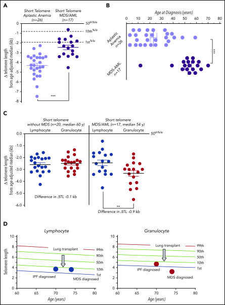
Short telomeres have been linked to cancer risk, yet other evidence supports them being tumor suppressive. Here, we report cancer outcomes in individuals with germline mutations in telomerase and other telomere-maintenance genes. Among 180 individuals evaluated in a hospital-based setting, 12.8% had cancer. Solid tumors were rare (2.8%); nearly all were young male DKC1 mutation carriers, and they were generally resectable with good short-term outcomes. Myelodysplastic syndrome (MDS) was most common, followed by acute myeloid leukemia (AML); they accounted for 75% of cancers. Age over 50 years was the biggest risk factor, and MDS/AML usually manifested with marrow hypoplasia and monosomy 7, but the somatic mutation landscape was indistinct from unselected patients. One- and 2-year survival were 61% and 39%, respectively, and two-thirds of MDS/AML patients died of pulmonary fibrosis and/or hepatopulmonary syndrome. In one-half of the cases, MDS/AML patients showed a recurrent peripheral blood pattern of acquired, granulocyte-specific telomere shortening. This attrition was absent in age-matched mutation carriers who did not have MDS/AML. We tested whether adult short telomere patients without MDS/AML also had evidence of clonal hematopoiesis of indeterminate potential-related mutations and found that 30% were affected. These patients also primarily suffered morbidity from pulmonary fibrosis during follow-up. Our data show that the Mendelian short telomere syndromes are associated with a relatively narrow cancer spectrum, primarily MDS and AML. They suggest that short telomere length is sufficient to drive premature age-related clonal hematopoiesis in these inherited disorders.


Conflict of interest statement

Conflict-of-interest disclosure: The authors declare no competing financial interests.

Figures

None
Graphical abstract
Figure 1.
Figure 1.
Prevalence and cancer phenotypes in patients with germline defects in telomere maintenance. (A) Telogram of lymphocyte telomere lengths of 176 short telomere syndrome patients relative to a nomogram of healthy controls with percentile lines labeled on the right. The mutant gene for each subject is color coded and the key is shown above the chart. Four patients included in this study were deceased prior to telomere-length measurement. (B) Telogram showing the patients with noncutaneous cancer diagnoses within the Johns Hopkins cohort of 176 patients in this study denoted in red; individuals without cancer are denoted in gray. One MDS/AML patient (among 23 with cancer) is not shown because of missing telomere-length data as explained in panel A. (C) Distribution of the 24 cancer types identified in 23 patients. (D) Telogram showing solid tumor and MDS/AML cancers by age and sex. The squares refer to male subjects and circles to female subects. DKC1 mutation carriers are annotated to show their young-onset disease and predilection to solid tumors.
Figure 2.
Figure 2.
Histopathologic and molecular characterization of short telomere MDS/AML. (A) Hypoplastic MDS (MDS with multilineage dysplasia; biopsy [hematoxylin-and-eosin stain (H&E); original magnification ×10]). (B) Multilineage dysplasia involving the myeloid (red arrows), erythroid (green arrow; aspirate [Wright-Giemsa stain; original magnification ×100]) and (C) megakaryocytic (yellow arrows; biopsy [H&E; original magnification ×40]) lineages. (D) MDS (MDS with ring sideroblasts and multilineage dysplasia; biopsy [H&E; original magnification ×10]). (E) Multilineage dysplasia involving the erythroid lineage (green arrows; aspirate [Wright-Giemsa stain; original magnification ×100]) with (F) abundant ring sideroblasts (aspirate [iron stain; original magnification ×100x]). Rare blasts (G, black arrow; Wright-Giemsa stain; original magnification ×100) are seen, and megakaryocytes (H) are also dysplastic (yellow arrows; H&E; original magnification ×40). (I) Hypoplastic AML (AML with myelodysplasia-related changes; biopsy [H&E; original magnification ×10]). Increased blasts (J, ∼70%) with background multilineage dysplasia involving the myeloid (K, red arrows) and erythroid (L, green arrows) lineages (aspirate [Wright-Giemsa stain; original magnification ×100]). (M) Hypoplastic AML (AML with myelodysplasia-related changes; biopsy [H&E; original magnification ×10]). Increased blasts (N; ∼50%) with background multilineage dysplasia involving the myeloid (O, red arrows) and erythroid (P, green arrow) lineages (aspirate [Wright-Giemsa stain; original magnification ×100]). (Q) Pie chart showing the distribution of karyotypes of 18 MDS/AML cases. Monosomy 7 abnormalities are listed on the left. (R) Somatic mutation profile of age-matched short telomere patients separated by those with (9 male/5 female) and without (11 male/9 female) MDS/AML. The age for each patient is included on top as is the presence of a monosomy 7 abnormality [−7, del(7q) or der(1;7)(q10;p10)]. All sequencing was performed on blood, except for the 2 AML cases (denoted as bolded ages) for which bone marrow was used. The ring sideroblasts (defined as >15% of cells examined) are denoted below the MDS/AML cases by an asterisk (*). Of note, another patient (of a total of 7) had ring sideroblasts but was not included in this analysis because of insufficient DNA. The respective germline mutations for each case are annotated in supplemental Figure 1A.
Figure 3.
Figure 3.
Telomere length discordance in short telomere patients with MDS/AML. (A) Mean of Δ telomere length from age-adjusted median in lymphocytes for patients with aplastic anemia and MDS/AML. (B) Age of diagnosis for patients with aplastic anemia and MDS/AML shows older age of onset for the latter group. (C) The degree of deviation from the age-adjusted median in lymphocytes and granulocytes in short telomere cases without (n = 20; median age, 60 years; range, 45-76 years; 13 male/7 female) and with MDS/AML (n = 17; median age, 54 years; range, 12-74 years; 11 male/6 female). Mean is graphed plus or minus standard error of the mean. **P = .001 (Mann-Whitney U test); ***P < .001 (Wilcoxon rank matched-pair test). (D) Longitudinal lymphocyte and granulocyte telomere lengths for 1 individual obtained at idiopathic pulmonary fibrosis (IPF) diagnosis and 4 years later at MDS diagnosis. The intervening age of lung transplant is graphically annotated by the arrow at age 72.
Figure 4.
Figure 4.
Treatment outcomes and clinical course of telomere-mediated MDS/AML. Swimmer plot of outcomes of 18 short telomere patients with MDS/AML clustered by co-occurrence of pulmonary fibrosis (PF). The age and treatment are shown to the right. Red bar shows time since diagnosis of MDS/AML, and purple indicates time since onset of pulmonary fibrosis symptoms. HSCT and lung transplant are graphically indicated along the timelines. The length of the bar represents follow-up through death or through last assessment. *This patient received standard acute lymphoblastic leukemia (ALL) therapy and therapy-related AML was diagnosed 8 months into the treatment, then he was treated with a mitoxantrone/etoposide salvage regimen. ***These 2 patients received standard intensive induction for high grade MDS and AML. ATG, antithymocyte globulin; Chemo, chemotherapy; CSA, cyclosporine; ESA, erythropoietin-stimulating agent; GCSF, granulocyte colony-stimulating factor; HMA, hypomethylating agent.

References

    1. McNally EJ, Luncsford PJ, Armanios M. Long telomeres and cancer risk: the price of cellular immortality. J Clin Invest. 2019;130(9):3474-3481. - PMC - PubMed
    1. Blasco MA, Lee HW, Hande MP, et al. . Telomere shortening and tumor formation by mouse cells lacking telomerase RNA. Cell. 1997;91(1):25-34. - PubMed
    1. Artandi SE, Chang S, Lee SL, et al. . Telomere dysfunction promotes non-reciprocal translocations and epithelial cancers in mice. Nature. 2000;406(6796):641-645. - PubMed
    1. Hao LY, Armanios M, Strong MA, et al. . Short telomeres, even in the presence of telomerase, limit tissue renewal capacity. Cell. 2005;123(6):1121-1131. - PubMed
    1. Armanios M, Alder JK, Parry EM, Karim B, Strong MA, Greider CW. Short telomeres are sufficient to cause the degenerative defects associated with aging. Am J Hum Genet. 2009;85(6):823-832. - PMC - PubMed

Publication types

MeSH terms