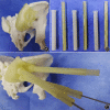
Combined 3D Printed Template to Guide Iliosacral Screw Insertion for Sacral Fracture and Dislocation: A Retrospective Analysis
- PMID: 32077257
- PMCID: PMC7031549
- DOI: 10.1111/os.12620
Combined 3D Printed Template to Guide Iliosacral Screw Insertion for Sacral Fracture and Dislocation: A Retrospective Analysis
Abstract
Objective: To evaluate the accuracy and safety of a combined 3D printed guide template (combined template) to assist iliosacral (IS) screw placement for sacral fracture and dislocation.
Methods: A total of 37 patients, 24 men and 13 women, age from 22 to 68 years old, diagnosed with a sacral fracture and dislocation were involved in this study for retrospective analysis from January 2016 to February 2018. There were 19 patients in the template group (42 screws) and 18 patients in the conventional group (31 screws). In the combined template group, IS screw placement was assisted by a combined 3D printed template; in the conventional group, the IS screws were placed freehand under fluoroscopy. The accuracy of the IS screw placement was evaluated by comparing the screw angle and the location of the screw entry point between the actual and the simulated screw in the combined template group. The safety of the IS screw placement was evaluated by comparing the quality of the reduction, the grading of the screws, the operation time, and radiation exposure times between groups.
Results: A total of 73 pedicle screws were placed in 37 patients: 42 screws (30 S1, 12 S2) in the combined template group and 31 screws (23 S1, 8 S2) in the conventional group. In the conventional group, 1 patient developed symptoms of L5 nerve stimulation. In the combined template group, the average operative time of each screw was 25.01 ± 2.90 min, with average radiation exposure times of 12.05 ± 4.00. In the conventional group, the average operative time of each screw was 46.24 ± 9.59 min, with an average radiation exposure time of 56.10 ± 6.75. There were significant differences in operation and radiation exposure times between groups. The rate of screw perforation was lower in the combined template group (2 of 42 screws, 0 at grade III and 2 at grade II) than in the conventional group (5 of 38 screws, 2 at grade III and 3 at grade III). In the combined template group, the mean distance between the entry points of the actual and simulated screws was 1.4 ± 0.9 mm, with a mean angle of deviation of 2.1° ± 1.6°. All patients were followed up once every 3 months and were followed for 3 to 12 months.
Conclusion: Using the combined template to assist with the insertion of IS screws delivered good accuracy, less fluoroscopy and shorter operation time, and avoided neurovascular injury as a result of screw malposition.
Keywords: 3D printing technology; Combined template; Iliosacral screw; Minimally invasive; Sacral fracture.
© 2020 The Authors. Orthopaedic Surgery published by Chinese Orthopaedic Association and John Wiley & Sons Australia, Ltd.
Figures


References
-
- Cooper KL, Beabout JW, Swee RG. Insufficiency fractures of the sacrum. Radiology, 1985, 156: 15–20. - PubMed
-
- Weber M, Hasler P, Gerber H. Insufficiency fractures of the sacrum: twenty cases and review of the literature. Spine, 1993, 18: 2507–2512. - PubMed
-
- Esses SI, Botsford DJ, Huler RJ, Rauschning W. Surgical anatomy of the sacrum. A guide for rational screw fixation. Spine, 1991, 16: 283–288. - PubMed
-
- Keating JF, Werier J, Blachut P, Broekhuyse H, Meek RN, O'Brien PJ. Early fixation of the vertically unstable pelvis: the role of IS screw fixation of the posterior lesion. J Orthop Trauma, 1999, 13: 107–113. - PubMed
MeSH terms
Grants and funding
LinkOut - more resources
Full Text Sources
Medical

