Review
. 2020 May;14(5):453-464.
doi: 10.1080/17476348.2020.1732822. Epub 2020 Feb 25.

Spinal cord injury and diaphragm neuromotor control

Affiliations
Review

Spinal cord injury and diaphragm neuromotor control

Matthew J Fogarty et al. Expert Rev Respir Med. 2020 May.

Abstract

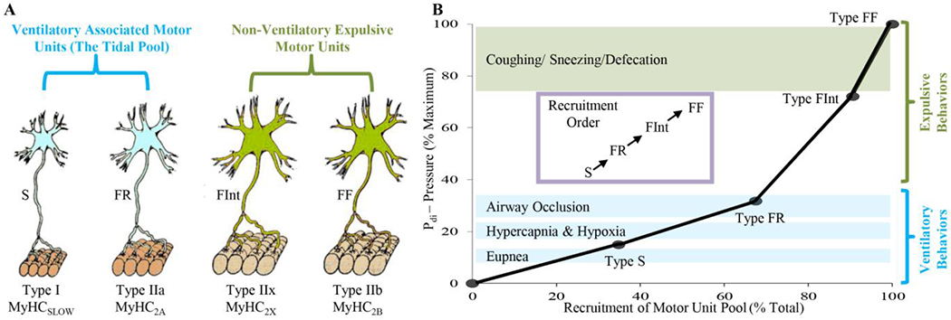
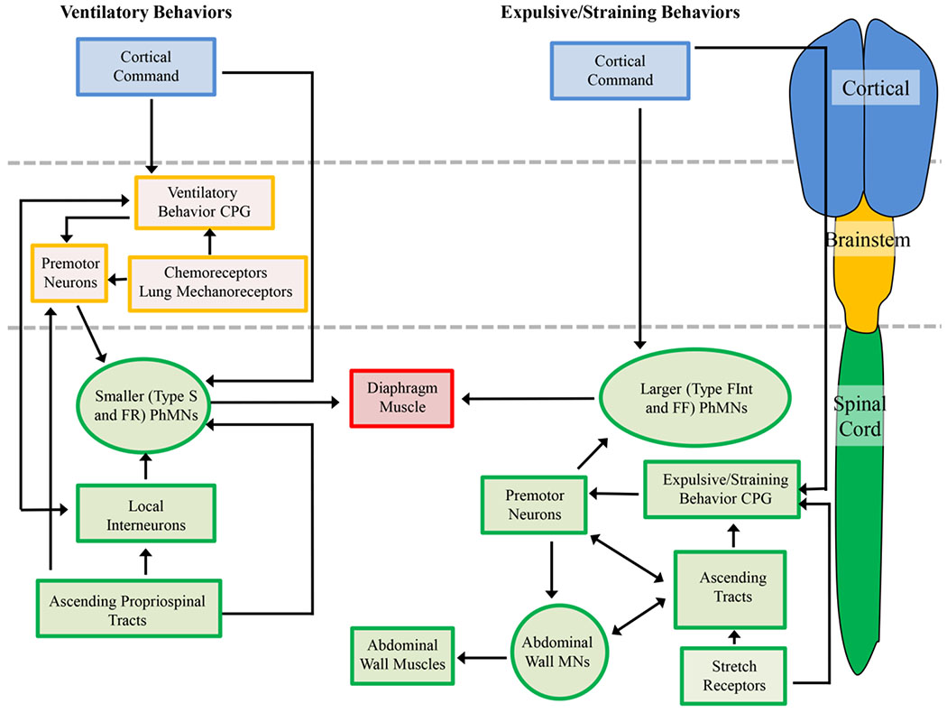
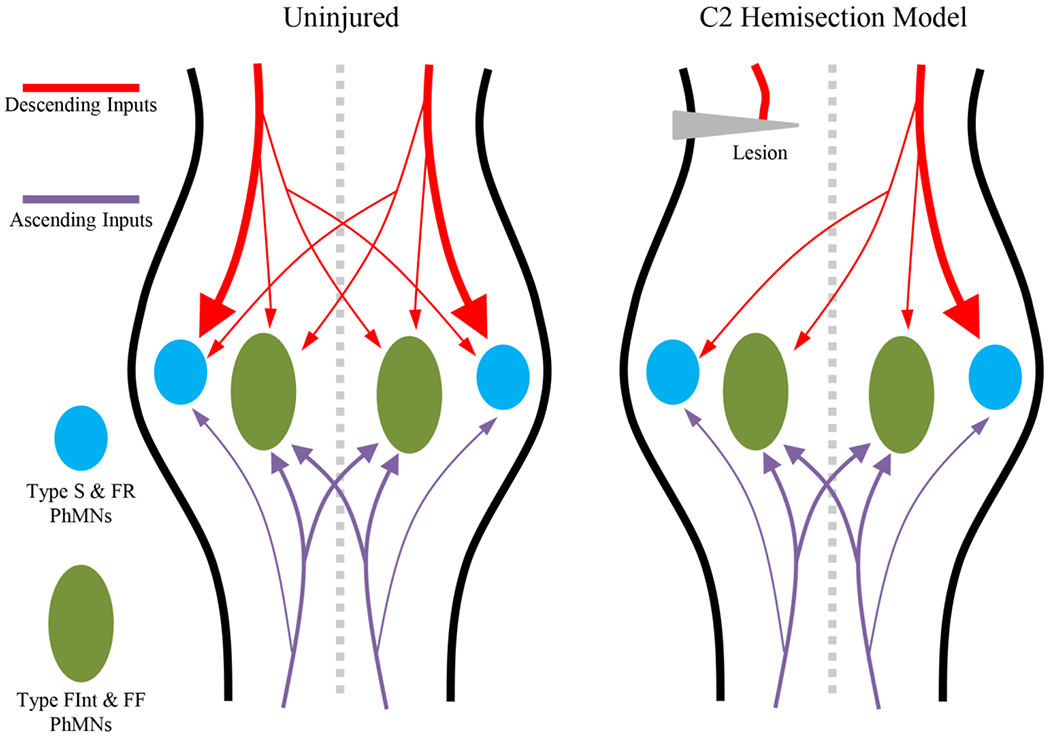
Introduction: Neuromotor control of diaphragm muscle and the recovery of diaphragm activity following spinal cord injury have been narrowly focused on ventilation. By contrast, the understanding of neuromotor control for non-ventilatory expulsive/straining maneuvers (including coughing, defecation, and parturition) is relatively impoverished. This variety of behaviors are achieved via the recruitment of the diverse array of motor units that comprise the diaphragm muscle.Areas covered: The neuromotor control of ventilatory and non-ventilatory behaviors in health and in the context of spinal cord injury is explored. Particular attention is played to the neuroplasticity of phrenic motor neurons in various models of cervical spinal cord injury.Expert opinion: There is a remarkable paucity in our understanding of neuromotor control of maneuvers in spinal cord injury patients. Dysfunction of these expulsive/straining maneuvers reduces patient quality of life and contributes to severe morbidity and mortality. As spinal cord injury patient life expectancies continue to climb steadily, a nexus of spinal cord injury and age-associated comorbidities are likely to occur. While current research remains concerned only with the minutiae of ventilation, the major functional deficits of this clinical cohort will persist intractably. We posit some future research directions to avoid this scenario.

Keywords: Phrenic motor neurons; contusion; hemisection; motor unit; neural circuit; recruitment; skeletal muscle.


Conflict of interest statement

Declaration of interest

The authors have no relevant affiliations or financial involvement with any organization or entity with a financial interest in or financial conflict with the subject matter or materials discussed in the manuscript. This includes employment, consultancies, honoraria, stock ownership or options, expert testimony, grants or patents received or pending, or royalties. The authors have nothing to disclose and declare no actual or perceived conflicts of interest.

Figures

Figure 1:
Figure 1:
Different diaphragm muscle motor unit types are distinguished by their intrinsic, mechanical, and fatigue properties, and are classified as type S, FR, FInt, and FF. Within an individual motor unit, all constituent muscle fibers exhibit homogeneous myosin heavy chain (MyHC) expression. In the diaphragm muscle of most species, type I and IIa muscle fibers have smaller cross-sectional areas than those of type IIx and/or IIb fibers. Forces produced by type I fibers are less than forces produced by type IIa fibers that are less than forces produced by IIx and/or IIb fibers. Recruitment of diaphragm muscle motor units is in an orderly fashion, necessary to accomplish a range of motor behaviors. Ventilation is accomplished by recruitment type S and FR motor units, whereas higher-force airway clearance behaviors and straining/expulsive manoeuvres require recruitment of more fatigueable type FInt and FF motor units.
Figure 2:
Figure 2:
Neuromotor control of diaphragm muscle ventilatory and expulsive/straining behaviors requires cortical (blue boxes), brainstem (orange boxes) and spinal cord (green boxes) centers. Ventilatory behaviors are the well characterized and require the recruitment of predominantly type S and FR motor units. Cortical pathways are able to modulate the eupnic rhythm by interactions with the ventilatory central pattern generator (CPG) or directly via synapses onto phrenic motor neurons (PhMNs). The ventilatory CPG activates brainstem premotor neurons that in turn innervate the PhMNs. Activity of PhMNs during ventilation is also modulated (directly and indirectly) by spinal cord ascending tracts and interneurons. Brainstem chemoreceptors and lung mechanoreceptors regulate the activity of premotor neurons, and act to increase premotor neuron discharge (and thus PhMN activity) during hypoxia/hypercapnia. In the case of expulsive/straining behaviors, the majority of control centers are located within the spinal cord, and recruitment of type FInt and FF motor units (higher-force producing units) is necessitated. Some cortical control of the PhMNs and spinal expulsive/straining CPG may be evident, but rectal and vaginal stretch receptors also elicit strong Pab generation. There may be shared spinal premotor neurons within the spinal cord for co-activations of PhMNs and abdominal muscle MNs, with a variety of ascending projections coordinating these activities.
Figure 3:
Figure 3:
The majority of phrenic motor neuron glutamatergic inputs are derived from descending (red) and ascending (purple) tracts. In the uninjured spinal cord, the majority of the bulbospinal descending inputs are distributed ipsilaterally to type S and FR phrenic motor neurons (PhMNs, blue), with small amounts of contralateral input to type S and FR PhMNs and a modicum of ipsi- and contralateral inputs to type FInt and FF motor units (green). Ascending inputs are primarily activated by co-contractions with abdominal muscles and both ipsi- and potentially contralateral inputs are predominantly on type FInt and FF PhMNs. Following unilateral C2 cervical hemisection, the inputs transected are the ipsilateral bulbospinal descending projections onto type S and FR PhMNs, accounting for impairments in ventilatory behaviors. In this model, the majority of inputs to type FInt and FF PhMNs remain, accounting for the preserved Pdimax.

Similar articles

Cited by

References

    1. Fogarty MJ, Sieck GC. Evolution and Functional Differentiation of the Diaphragm Muscle of Mammals. Compr Physiol. 2019;9(2):715–766. - PMC - PubMed
    2. * This review is contains a synthesis of the best information available regarding the ventilatory and non-ventilatory behaviours of the diaphragm muscle.

    1. Fogarty MJ, Mantilla CB, Sieck GC. Breathing: Motor Control of Diaphragm Muscle. Physiology (Bethesda). 2018. March 1;33(2):113–126. doi: 10.1152/physiol.00002.2018. - DOI - PMC - PubMed
    1. Lisboa C, Pare PD, Pertuze J, et al. Inspiratory muscle function in unilateral diaphragmatic paralysis. Am Rev Respir Dis. 1986. September;134(3):488–92. doi: 10.1164/arrd.1986.134.3.488. - DOI - PubMed
    1. Khurram OU, Fogarty MJ, Sarrafian TL, et al. Impact of aging on diaphragm muscle function in male and female Fischer 344 rats. Physiol Rep. 2018. July;6(13):e13786. doi: 10.14814/phy2.13786. - DOI - PMC - PubMed
    1. Sieck GC, Fournier M. Diaphragm motor unit recruitment during ventilatory and nonventilatory behaviors. J Appl Physiol. 1989;66(6):2539–2545. - PubMed

Publication types