

Improving Oral Cancer Outcomes with Imaging and Artificial Intelligence
- PMID: 32077795
- PMCID: PMC7036512
- DOI: 10.1177/0022034520902128
Improving Oral Cancer Outcomes with Imaging and Artificial Intelligence
Abstract
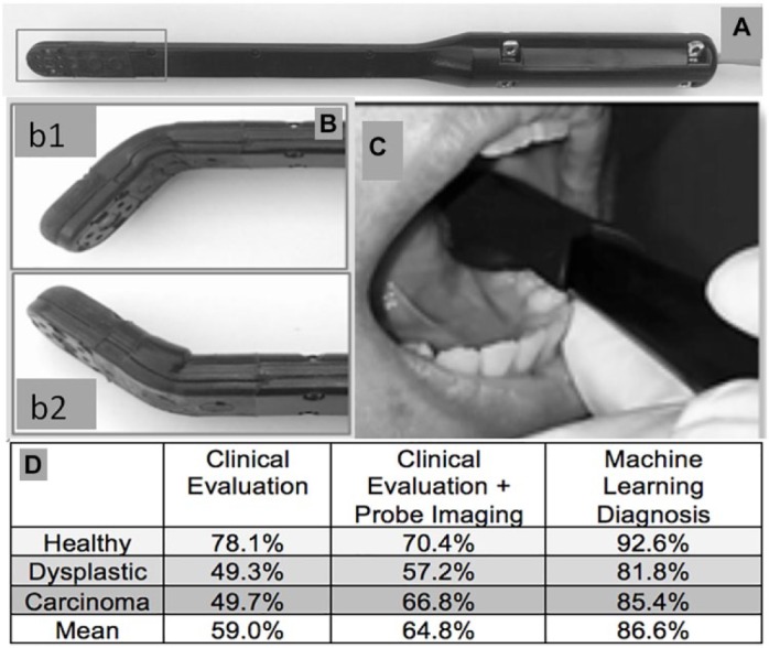
Early diagnosis is the most important determinant of oral and oropharyngeal squamous cell carcinoma (OPSCC) outcomes, yet most of these cancers are detected late, when outcomes are poor. Typically, nonspecialists such as dentists screen for oral cancer risk, and then they refer high-risk patients to specialists for biopsy-based diagnosis. Because the clinical appearance of oral mucosal lesions is not an adequate indicator of their diagnosis, status, or risk level, this initial triage process is inaccurate, with poor sensitivity and specificity. The objective of this study is to provide an overview of emerging optical imaging modalities and novel artificial intelligence-based approaches, as well as to evaluate their individual and combined utility and implications for improving oral cancer detection and outcomes. The principles of image-based approaches to detecting oral cancer are placed within the context of clinical needs and parameters. A brief overview of artificial intelligence approaches and algorithms is presented, and studies that use these 2 approaches singly and together are cited and evaluated. In recent years, a range of novel imaging modalities has been investigated for their applicability to improving oral cancer outcomes, yet none of them have found widespread adoption or significantly affected clinical practice or outcomes. Artificial intelligence approaches are beginning to have considerable impact in improving diagnostic accuracy in some fields of medicine, but to date, only limited studies apply to oral cancer. These studies demonstrate that artificial intelligence approaches combined with imaging can have considerable impact on oral cancer outcomes, with applications ranging from low-cost screening with smartphone-based probes to algorithm-guided detection of oral lesion heterogeneity and margins using optical coherence tomography. Combined imaging and artificial intelligence approaches can improve oral cancer outcomes through improved detection and diagnosis.
Keywords: dentists; diagnosis; machine intelligence; medicine; oral neoplasms; screening.
Conflict of interest statement
The authors declare no potential conflicts of interest with respect to the authorship and/or publication of this article.
Figures
References
-
- Alston PA, Knapp J, Luomanen JC. 2014. Who will tend the dental safety net? J Calif Dent Assoc. 42(2):112–118. - PubMed
-
- Amarasinghe HK, Usgodaarachchi US, Johnson NW, Lalloo R, Warnakulasuriya S. 2010. Public awareness of oral cancer, of oral potentially malignant disorders and of their risk factors in some rural populations in Sri Lanka. Community Dent Oral Epidemiol. 38(6):540–548. - PubMed
-
- American Academy of Oral Medicine (AAOM). 2019. Subject: oral cancer screening [accessed 2019 June 4]. https://www.aaom.com/clinical-practice-statement-oral-cancer-screening
-
- American Cancer Society (ACS). 2018. Cancer facts and figures. Atlanta (GA): ACS.
