Review
. 2020 Feb 16;20(4):1072.
doi: 10.3390/s20041072.

Applications of Graphene Quantum Dots in Biomedical Sensors

Affiliations
Review

Applications of Graphene Quantum Dots in Biomedical Sensors

Bhargav D Mansuriya et al. Sensors (Basel). .

Abstract

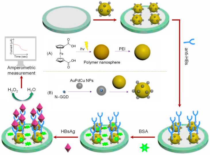
Due to the proliferative cancer rates, cardiovascular diseases, neurodegenerative disorders, autoimmune diseases and a plethora of infections across the globe, it is essential to introduce strategies that can rapidly and specifically detect the ultralow concentrations of relevant biomarkers, pathogens, toxins and pharmaceuticals in biological matrices. Considering these pathophysiologies, various research works have become necessary to fabricate biosensors for their early diagnosis and treatment, using nanomaterials like quantum dots (QDs). These nanomaterials effectively ameliorate the sensor performance with respect to their reproducibility, selectivity as well as sensitivity. In particular, graphene quantum dots (GQDs), which are ideally graphene fragments of nanometer size, constitute discrete features such as acting as attractive fluorophores and excellent electro-catalysts owing to their photo-stability, water-solubility, biocompatibility, non-toxicity and lucrativeness that make them favorable candidates for a wide range of novel biomedical applications. Herein, we reviewed about 300 biomedical studies reported over the last five years which entail the state of art as well as some pioneering ideas with respect to the prominent role of GQDs, especially in the development of optical, electrochemical and photoelectrochemical biosensors. Additionally, we outline the ideal properties of GQDs, their eclectic methods of synthesis, and the general principle behind several biosensing techniques.

Keywords: biomedical applications; biosensors; electrochemical sensors; graphene quantum dots (GQDs); nanomaterials; optical sensors; photoelectrochemical sensors.


Conflict of interest statement

The authors declare no conflict of interest.

Figures

Figure 1
Figure 1
The chemical structure of graphene quantum dots (GQDs) and their biomedical applications in sensor development.
Figure 2
Figure 2
Schematic representation of a fluorescent assay for DNA methyltransferase activity [95].
Figure 3
Figure 3
The detection principle of the aptamer/Fe3O4/GQDs/MoS2-based nanosurface energy transfer biosensor for sensing circulating tumor cells (CTCs).
Figure 4
Figure 4
Detection mechanism of an enzyme based glucose-sensor. Adapted from Wang et al. [114].
Figure 5
Figure 5
Schematic representation of: (A) Enzyme-based photoluminescence (PL) sensor for dichlorvos [156]; (B) MIP-based PL sensor for tributyltin [157].
Figure 6
Figure 6
A GQD sensor based on PL for screening protein kinase activity [162].
Figure 7
Figure 7
Schematic illustration of various transition metal ions and their influences on the PL of GQDs [168].
Figure 8
Figure 8
Construction of MIP@N‒GQDs based chemiluminescence (CL) sensor for the quantification of doxorubicin [178]. Reproduced by permission of The Royal Society of Chemistry. APTES: Aminopropyltriethoxysilane; TEOS: Tetraethoxysilane.
Figure 9
Figure 9
Schematic illustration of CL-based GQD sensor for CA-125 determination [181]. Reproduced by permission of The Royal Society of Chemistry.
Figure 10
Figure 10
A step-wise developmental procedure of GQD-based sandwich-assay for carbohydrate antigen (CA) 199 [191].
Figure 11
Figure 11
Detection strategy of nitroaniline by N‒GQDs and chitosan functionalized glassy carbon electrode (GCE) [207].
Figure 12
Figure 12
The mechanism involved in detecting DNA by GQD electrochemiluminescence (ECL) assembled with the multiple cycling amplification method [212].
Figure 13
Figure 13
DNA detection strategy of GQD-based fluorescence resonance energy transfer (FRET) nanosensor [224].
Figure 14
Figure 14
Biosensing system based on FRET change between GQDs and pyrene-modified molecular beacon probes (py-MBs) for determining miRNAs [236].
Figure 15
Figure 15
(A) Cyclic voltammetry (CV): potential vs. current profile with reference to a GQD-implanted electrode surface, where its electrochemical response changes according to the specific bio-recognition event. (B) Square wave voltammetry (SWV): potential vs. time profile, where t: time required for each pulse, f: frequency of each pulse, E1 and E2: initial and final pulse, respectively. (C) Target biomolecule detection by GQD-coated electrode based on SWV, where the current changes in direct correlation with an occupancy of a specific receptor. (D) Ideal curves observed with amperometric measurements for varied concentrations of target biomolecules. (E) Electron impedance spectroscopy (EIS): variation in impedance due to the modification of electrode surface and subsequent introduction of a target analyte (i.e., Nyquist plot). Adapted from Mansuriya and Altintas [79].
Figure 16
Figure 16
An example of a GQD-based electrochemical DNA biosensor.
Figure 17
Figure 17
(A) Casting procedure of GQD@VMSF (vertically-oriented mesoporous silica-nanochannel film) modified electrode via electrophoresis. (B) Quantification of Cu2+, Hg2+ and Cd2+ ions as well as dopamine. Adapted from Lu et al. [275].
Figure 18
Figure 18
Various steps tangled in designing a GQD-based antibody sensor for tracking human chorionic gonadotropin (HCG) levels [79].
Figure 19
Figure 19
Experimental steps to fabricate a GCE for detecting breast cancer [79].
Figure 20
Figure 20
Schematic presentation of a voltammetric antibody sensor for cTnI [79].
Figure 21
Figure 21
Set-up of an electrochemical and fluorescence based DNA-sensor for APO e4 DNA [306].
Figure 22
Figure 22
(A) Preparatory steps for PtPd/N‒GQDs/Au nanohybrid. (B) Schematic portrait of a label-free amperometric antibody-sensor for CEA determination. Adapted from Mansuriya and Altintas [79].
Figure 23
Figure 23
Schematic illustration of a GQD-based antibody sensor for Y. enterecolitica quantification [67].
Figure 24
Figure 24
A picturized illustration of a label-free amperometric antibody-sensor for HBV virus and synthesis of: (A) polyethylenimine (PEI)-modified polymer nanospheres (PS); (B) N‒GQDs/AuPdCu @ PS. Adapted from Mansuriya and Altintas [79].
Figure 25
Figure 25
(A) Preparation of GQDs. (B) Immunosensing platform for AFB1 using GQD-functionalized indium tin oxide (ITO)-coated glass electrode [79].
Figure 26
Figure 26
A label-free impedimetric sensing system for CEA. Adapted from Mansuriya and Altintas [79].
Figure 27
Figure 27
Detection of S. typhimurium by an impedimetric antibody sensor, followed by its response towards antibiotics, where (a) increased impedance (b) decreased impedance [79].
Figure 28
Figure 28
Fabrication of an N,S‒GQD-based photoelectrochemical (PEC) antibody-sensor for cTnI monitoring [353].
Figure 29
Figure 29
Design of a PEC aptasensor for zeatin determination using GQDs, AuNPs, g-C3N4 and DNA biotin-labeled aptamer [359].
Figure 30
Figure 30
Schematic representation of an surface plasmon resonance (SPR)sensor. Adapted from Ramdzan et al. [361].
Figure 31
Figure 31
The mechanism of RLS of GQDs for detecting mutant DNA based on catalytic hairpin assembly amplification approach [363].
Figure 32
Figure 32
Preparation of GQDs/AgNPs nanocomposite for colorimetric detection of H2O2 and glucose. Adapted from Chen et al. [369].

References

    1. Bray F., Ferlay J., Soerjomataram I., Siegel R.L., Torre L.A., Jemal A. Global cancer statistics 2018: GLOBOCAN estimates of incidence and mortality worldwide for 36 cancers in 185 countries. CA Cancer J. Clin. 2018;68:394–424. doi: 10.3322/caac.21492. - DOI - PubMed
    1. Stewart J., Manmathan G., Wilkinson P. Primary prevention of cardiovascular disease: A review of contemporary guidance and literature. JRSM Cardiovasc. Dis. 2017;6:204800401668721. doi: 10.1177/2048004016687211. - DOI - PMC - PubMed
    1. Ho K.J. Cardiovascular diseases. Nutr. Asp. Aging. 2018;2:75–100.
    1. Clark L.C., Lyons C. Electrode Systems for Continuous Monitoring in Cardiovascular Surgery. Ann. N. Y. Acad. Sci. 1962;102:29–45. doi: 10.1111/j.1749-6632.1962.tb13623.x. - DOI - PubMed
    1. Sharma T.K., Ramanathan R., Rakwal R., Agrawal G.K., Bansal V. Moving forward in plant food safety and security through NanoBioSensors: Adopt or adapt biomedical technologies? Proteomics. 2015:1680–1692. doi: 10.1002/pmic.201400503. - DOI - PubMed

LinkOut - more resources