Review
. 2020 Feb 17;9(2):542.
doi: 10.3390/jcm9020542.

Progress in Microneedle-Mediated Protein Delivery

Affiliations
Review

Progress in Microneedle-Mediated Protein Delivery

Rezvan Jamaledin et al. J Clin Med. .

Abstract

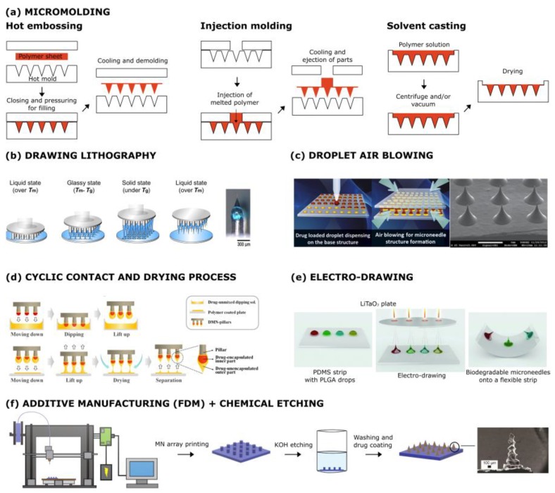
The growing demand for patient-compliance therapies in recent years has led to the development of transdermal drug delivery, which possesses several advantages compared with conventional methods. Delivering protein through the skin by transdermal patches is extremely difficult due to the presence of the stratum corneum which restricts the application to lipophilic drugs with relatively low molecular weight. To overcome these limitations, microneedle (MN) patches, consisting of micro/miniature-sized needles, are a promising tool to perforate the stratum corneum and to release drugs and proteins into the dermis following a non-invasive route. This review investigates the fabrication methods, protein delivery, and translational considerations for the industrial scaling-up of polymeric MNs for dermal protein delivery.

Keywords: antigen delivery; drug delivery; microneedles; protein; transdermal.


Conflict of interest statement

The authors declare no conflict of interest.

Figures

Figure 1
Figure 1
Microneedle (MN) manufacturing methods. (a) Micromolding: The mold with the desired MN structures can be filled with polymers by hot embossing, injection molding, or solvent casting. (b) Drawing lithography: The polymer is melted, dispensed on a fixed plate, and elongated by pillars in the upper-moving plate. (c) Droplet air blowing: Two plates, with polymer drops within, are contacted and then moved. When the final distance between the plates is reached, the polymer is hardened by means of air blowing. (d) Cyclic contact and drying: Pillars are repeatedly contacted with a drug-polymer solution, lifted, and dried with air blowing. (e) Electro-drawing: A thermal stimulus is applied to a pyroelectric crystal, generating an electric field which drives the microneedle drawing process. (f) Fused deposition modeling (FDM) of biodegradable polymer MNs: FDM is followed by KOH etching to improve feature size. Reprinted with modification from [36,37,38,39,40,41].
Figure 2
Figure 2
Yield strength vs. Young’s modulus of different materials used for the fabrication of microneedles. Reprinted with permission from [64].
Figure 3
Figure 3
Assembly method for immune polyelectrolyte multilayers on microneedle arrays to enhance cancer vaccination. Reprinted with permission from [102]. Immune polyelectrolyte multilayers (iPEM), cytosine triphosphate deoxynucleotide guanine triphosphate deoxynucleotide (CpG).
Figure 4
Figure 4
Functional test of MNs in a full-thickness human skin model. The pictures in (AC) refer to the encapsulating enzyme in the tip; (DF) refer to enzyme-encapsulated microparticles. A D Histological images after 48 h; black asterisks indicate the polyvinyl pyrrolidone (PVP) polymer remained after removing the patch (scale bar = 100 µm). B E Stereomicroscopic images of Endo-Human Skin Equivalent (Endo-HSE) (histological) 48 h after indentation (scale bar = 500 µm). The inserts of the stereomicroscopic images are the schematic representation of the methods used to calculate the diffusive radius reported in the x-axis of the successive graphs. (C,F): The graphs plot, at three time points, the pixel intensity as it corresponds to the concentration of the substrate oxidation product diffusing into the extracellular matrix vs. the radius of the diffusion pattern. Reprinted with permission from [62].
Figure 5
Figure 5
Schematic view of composite microparticle and bulk poly(lactic-co-glycolic) acid (PLGA) tip MN fabrication. Molds were first filled with PLGA microparticles (1). PLGA microparticles were then either dried in mold cavities (2a) or fused at a high temperature to create a solid tip (2b). Concentrated aq. poly(acrylic acid) solution was then centrifuged onto the filled molds to create a supportive matrix (3a) or pedestal (3b) for rapid dissolution in vivo. After drying, MNs were removed from molds (4a, 4b). Reprinted with permission from [117]. poly(lactic-co-glycolic) acid (PLGA), MP (microparticle), PDMS (polydimethylsiloxane), PAA poly(acrylic acid).
Figure 6
Figure 6
Fabrication process of phase transition patches. (A) The MNs absorb the interstitial fluid (IF) from the dermis layer to convert from a hard, glassy state to a hydrogel state to allow the preloaded insulin to release to the bodily fluid in the dermis layer. (B) The microneedle matrix of phase-transition microneedles (PTM) is cross-linked through microcrystalline domains as the cross-linking junctions via a freeze–thaw treatment to avoid dissolution, while that of hydrogel forming is cross-linked through covalent bands as the cross-linking junctions via a chemical reaction. Therefore, insulin can be loaded in the needle tips of PTM to achieve a relative bioavailability of 20%, while insulin has to be loaded at the back of the microneedle array of hydrogel-forming microneedles (HFMs), leading to a bioavailability of less than 1% due to the extended diffusion pathway. (C) The PTM patch may be fabricated using a scalable process comprising a sequence of simple unit operations involving the circulation of the molds in the production line and sterilization of the final product by steaming in oxirane vapor. Reprinted with permission from [123].
Figure 7
Figure 7
Bioactive glass nanoparticles (BGN) can be manufactured by BGN soles through gelation under high temperatures. These BGNs were filled with GOx/catalase (CAT) inside their pores, and then the NPs were coated by ZnO quantum dots (QDs). As BGLs grow, the pH drops to a value lower than 5.5 as a result of the reaction occurring by GOx. ZnO QDs are dissolved under this low pH, which disintegrates the BGN, and thus, insulin is free to be released from the disassembled particle. The catalase (CAT) enzyme is responsible for reducing the harm caused by H2O2 on the surrounding tissue. Reprinted with permission from [134].
Figure 8
Figure 8
(A) Live glucose-responsive design based on the diffusion of glucose in cross-linked hyaluronic acid and stimulation of cells. Unfortunately, no significant release was observed at different glucose concentrations due to the low glucose diffusion in MNs. (B) Living-synthetic responsive system with amplified glucose levels, aided by nanovesicles containing three enzymes. After sensing local hyperglycemia, the reaction by glucose oxidase occurs, which results in a decrease in pH and leads to low O2 levels. Thus, hypoxia-sensitive nanoparticles dissociate, resulting in the release of all enzymes. Released α-amylase and glucoamylase convert α-amylose to glucose in two separate steps. Higher amounts of glucose diffuse and make the living part of the system secrete insulin effectively. Reprinted with permission from [135].

References

    1. Leader B., Baca Q.J., Golan D.E. Protein therapeutics: A summary and pharmacological classification. Nat. Rev. Drug Discov. 2008;7:21. doi: 10.1038/nrd2399. - DOI - PubMed
    1. Rawat A., Burgess D.J. Parenteral Delivery of Peptides and Proteins, in Biodrug Delivery Systems. CRC Press; Boca Raton, FL, USA: 2016. pp. 66–84.
    1. Patel A., Cholkar K., Mitra A.K. Recent developments in protein and peptide parenteral delivery approaches. Ther. Deliv. 2014;5:337–365. doi: 10.4155/tde.14.5. - DOI - PMC - PubMed
    1. Lundquist P., Artursson P. Oral absorption of peptides and nanoparticles across the human intestine: Opportunities, limitations and studies in human tissues. Adv. Drug Deliv. Rev. 2016;106:256–276. doi: 10.1016/j.addr.2016.07.007. - DOI - PubMed
    1. Al-Mayahy M.H., Sabri A.H., Rutland C.S., Holmes A., McKenna J., Marlow M., Scurr D.J. Insight into imiquimod skin permeation and increased delivery using microneedle pre-treatment. Eur. J. Pharm. Biopharm. 2019;139:33–43. doi: 10.1016/j.ejpb.2019.02.006. - DOI - PubMed

LinkOut - more resources