

Role of Signal Transduction Pathways and Transcription Factors in Cartilage and Joint Diseases
- PMID: 32079226
- PMCID: PMC7072930
- DOI: 10.3390/ijms21041340
Role of Signal Transduction Pathways and Transcription Factors in Cartilage and Joint Diseases
Abstract
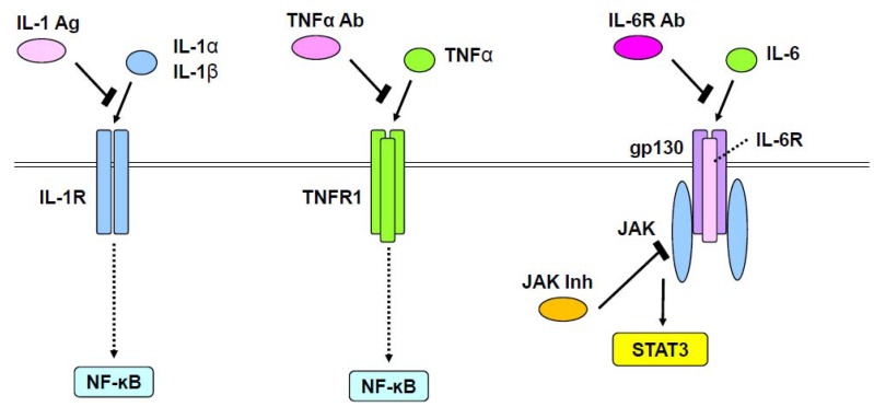
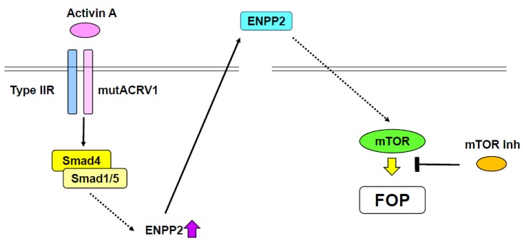
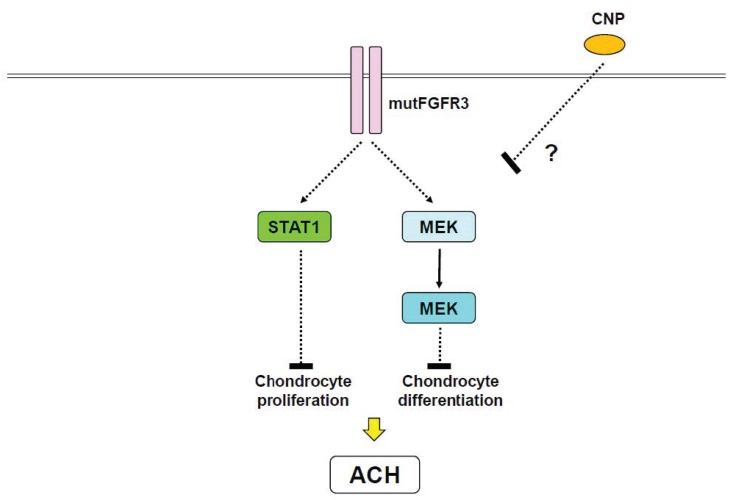
: Osteoarthritis and rheumatoid arthritis are common cartilage and joint diseases that globally affect more than 200 million and 20 million people, respectively. Several transcription factors have been implicated in the onset and progression of osteoarthritis, including Runx2, C/EBPβ, HIF2α, Sox4, and Sox11. Interleukin-1 β (IL-1β) leads to osteoarthritis through NF-ĸB, IκBζ, and the Zn2+-ZIP8-MTF1 axis. IL-1, IL-6, and tumor necrosis factor α (TNFα) play a major pathological role in rheumatoid arthritis through NF-ĸB and JAK/STAT pathways. Indeed, inhibitory reagents for IL-1, IL-6, and TNFα provide clinical benefits for rheumatoid arthritis patients. Several growth factors, such as bone morphogenetic protein (BMP), fibroblast growth factor (FGF), parathyroid hormone-related protein (PTHrP), and Indian hedgehog, play roles in regulating chondrocyte proliferation and differentiation. Disruption and excess of these signaling pathways cause genetic disorders in cartilage and skeletal tissues. Fibrodysplasia ossificans progressive, an autosomal genetic disorder characterized by ectopic ossification, is induced by mutant ACVR1. Mechanistic target of rapamycin kinase (mTOR) inhibitors can prevent ectopic ossification induced by ACVR1 mutations. C-type natriuretic peptide is currently the most promising therapy for achondroplasia and related autosomal genetic diseases that manifest severe dwarfism. In these ways, investigation of cartilage and chondrocyte diseases at molecular and cellular levels has enlightened the development of effective therapies. Thus, identification of signaling pathways and transcription factors implicated in these diseases is important.
Keywords: achondroplasia; fibrodysplasia ossificans progressive; osteoarthritis; rheumatoid arthritis.
Conflict of interest statement
The authors declare no conflict of interest.
Figures
References
Publication types
MeSH terms
Substances
LinkOut - more resources
Full Text Sources
Other Literature Sources
Medical
Research Materials
Miscellaneous
