Review
. 2020 Feb 17;12(2):464.
doi: 10.3390/polym12020464.

Protein-Polysaccharide Composite Materials: Fabrication and Applications

Affiliations
Review

Protein-Polysaccharide Composite Materials: Fabrication and Applications

Elizabeth J Bealer et al. Polymers (Basel). .

Abstract

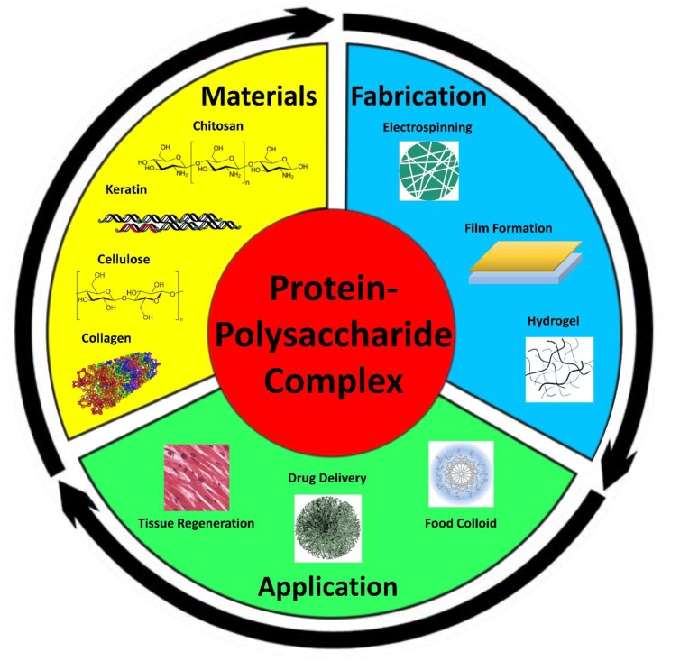
Protein-polysaccharide composites have been known to show a wide range of applications in biomedical and green chemical fields. These composites have been fabricated into a variety of forms, such as films, fibers, particles, and gels, dependent upon their specific applications. Post treatments of these composites, such as enhancing chemical and physical changes, have been shown to favorably alter their structure and properties, allowing for specificity of medical treatments. Protein-polysaccharide composite materials introduce many opportunities to improve biological functions and contemporary technological functions. Current applications involving the replication of artificial tissues in tissue regeneration, wound therapy, effective drug delivery systems, and food colloids have benefited from protein-polysaccharide composite materials. Although there is limited research on the development of protein-polysaccharide composites, studies have proven their effectiveness and advantages amongst multiple fields. This review aims to provide insight on the elements of protein-polysaccharide complexes, how they are formed, and how they can be applied in modern material science and engineering.

Keywords: composite material; drug delivery and nanomedicine; health and nutrition; protein and polysaccharide; tissue regeneration; water treatment.


Conflict of interest statement

The authors declare no conflict of interest.

Figures

Figure 1
Figure 1
Protein–polysaccharide composite materials can be fabricated from a variety of protein and polysaccharide sources. These complex materials can then be processed into variable shapes with unique properties for a multitude of applications.
Figure 2
Figure 2
Complex coacervation process showing the combination of protein and polysaccharide (reproduced with permission from [84,85]. Copyright Elsevier, 2010 and 2017).
Figure 3
Figure 3
Schematic of electrospinning systems with respective orientation and SEM images of the fibers for the different protein–polysaccharide systems (left: collagen–chitosan composites; right: amaranth protein–pullulan blends) (reproduced with permission from [86,87], Copyright, Elsevier 2010 and 2013).
Figure 4
Figure 4
Formation of protein–polysaccharide gel complexes at different stages (reproduced with permission from [102] Copyright Elsevier, 2017).
Figure 5
Figure 5
Ternary-phase diagram of a protein/polysaccharide solvent system.
Figure 6
Figure 6
(AC) SEM images (scale: 100 μm) of the different protein–polysaccharide materials: (A) Alginate/gelatin (B) Alginate/collagen (C) Decellularized porcine myocardium. (D) Mechanical characterization of these protein–polysaccharide materials showing different storage and loss modulus. (EJ) Fluorescence micrographs displaying cardiomyocytes response to alginate/gelatin scaffolds at different days under static and dynamic conditions (AJ reproduced with permission from [121], Copyright Wiley, 2017).
Figure 7
Figure 7
Compressive strength of the different protein–polysaccharide composites: Bulk composite of nHCP/CG (nHA/chitosan–pectin/chitosan–gelatin) is similar to that of cancellous bone (reproduced with permission from [123], copyright Elsevier, 2011).
Figure 8
Figure 8
(A) The encapsulating capacity of different hydrogel/micelle systems. (B) In vitro drug release profiles of micelles and different hydrogel/micelle systems. (C) The drug release profiles of hydrogel/micelle = 1:1 and (D) hydrogel/micelle = 3:1 systems in simulated gastric fluid (SGF), simulated small intestinal fluid (SIF), and simulated colonic fluid (SCF), individually (AD reproduced with permission from [133], copyright Elsevier, 2018).
Figure 9
Figure 9
Novel applications of protein–polysaccharide composite materials: A) SEM images of the silymarin–zein nanoparticle/bacterial cellulose nanofibers used in a wound dressing application; B) Swelling ratio and release of protein from chitosan–pectin particles with change of the Ca2+ concentration in acidic solution; C) AFM 3 × 3 μm image of protein–polysaccharide (β-lactoglobulin with high methoxyl pectin) particles that were heat treated at pH 4.75, 83 °C (reproduced with permission from [114,131,144]. Copyright Elsevier, 2000, 2011, 2018).

References

    1. Azevedo M., Reis R., Claase M., Grijpma D., Feijen J. Development and properties of polycaprolactone/hydroxyapatite composite biomaterials. J. Mater. Sci. Mater. Med. 2003;14:103–107. doi: 10.1023/A:1022051326282. - DOI - PubMed
    1. Zheng Y., Monty J., Linhardt R.J. Polysaccharide-based nanocomposites and their applications. Carbohydr. Res. 2015;405:23–32. doi: 10.1016/j.carres.2014.07.016. - DOI - PMC - PubMed
    1. Suh J.-K.F., Matthew H.W. Application of chitosan-based polysaccharide biomaterials in cartilage tissue engineering: A review. Biomaterials. 2000;21:2589–2598. - PubMed
    1. Miao J., Pangule R.C., Paskaleva E.E., Hwang E.E., Kane R.S., Linhardt R.J., Dordick J.S. Lysostaphin-functionalized cellulose fibers with antistaphylococcal activity for wound healing applications. Biomaterials. 2011;32:9557–9567. doi: 10.1016/j.biomaterials.2011.08.080. - DOI - PubMed
    1. Liu L., Liu L., Liu C.-K., Fishman M.L., Hicks K.B. Composite films from pectin and fish skin gelatin or soybean flour protein. J. Agric. Food Chem. 2007;55:2349–2355. doi: 10.1021/jf062612u. - DOI - PubMed

LinkOut - more resources