. 2020 Apr 14;133(7):jcs241513.
doi: 10.1242/jcs.241513.

Regulation of intrinsic polarity establishment by a differentiation-type MAPK pathway in S. cerevisiae

Affiliations

Regulation of intrinsic polarity establishment by a differentiation-type MAPK pathway in S. cerevisiae

Aditi Prabhakar et al. J Cell Sci. .

Abstract

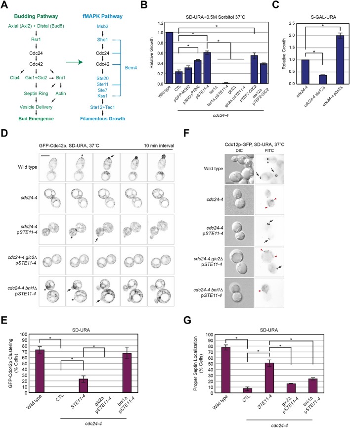
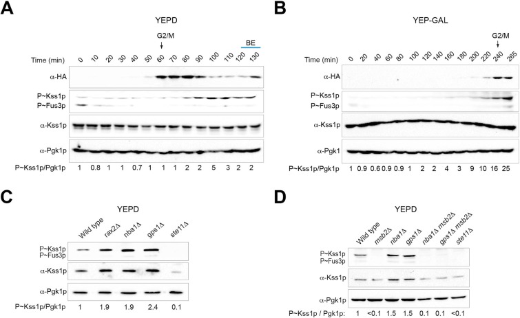
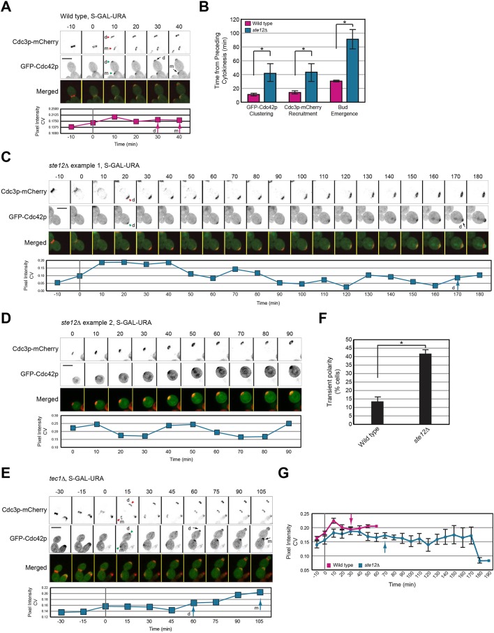
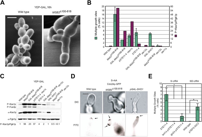
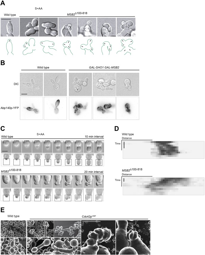
All cells establish and maintain an axis of polarity that is critical for cell shape and progression through the cell cycle. A well-studied example of polarity establishment is bud emergence in the yeast Saccharomyces cerevisiae, which is controlled by the Rho GTPase Cdc42p. The prevailing view of bud emergence does not account for regulation by extrinsic cues. Here, we show that the filamentous growth mitogen activated protein kinase (fMAPK) pathway regulates bud emergence under nutrient-limiting conditions. The fMAPK pathway regulated the expression of polarity targets including the gene encoding a direct effector of Cdc42p, Gic2p. The fMAPK pathway also stimulated GTP-Cdc42p levels, which is a critical determinant of polarity establishment. The fMAPK pathway activity was spatially restricted to bud sites and active during the period of the cell cycle leading up to bud emergence. Time-lapse fluorescence microscopy showed that the fMAPK pathway stimulated the rate of bud emergence during filamentous growth. Unregulated activation of the fMAPK pathway induced multiple rounds of symmetry breaking inside the growing bud. Collectively, our findings identify a new regulatory aspect of bud emergence that sensitizes this essential cellular process to external cues.

Keywords: Bud emergence; Cdc42p; MAPK; Polarity establishment; Pseudohyphal growth; Symmetry breaking.


Conflict of interest statement

Competing interestsThe authors declare no competing or financial interests.

Figures

Fig. 1.
Fig. 1.
Role of the fMAPK pathway in rescuing the growth defect of the cdc24-4 mutant. (A) Pathways that regulate bud emergence (green) and filamentous growth (blue). Cdc24p and Cdc42p regulate both pathways. Not all proteins are shown. (B) Growth of the indicated strains relative to wild-type cells. pGFP-MSB2, pSHO1P120L, pSTE11-4 and pTEF2-GIC2 were expressed from plasmids (see Table S2) in the indicated strains (see Table S1). Error bars show the standard error of the mean (s.e.m.) for three separate trials. *P<0.01. CTL, plasmid pRS316. (C) Growth of strains relative to the cdc24-4 mutant. This experiment was performed in galactose (GAL) medium, which also compromised the viability of the cdc24-4 mutant, because the pbs2Δ mutant has a growth defect at 37°C (Winkler et al., 2002). (D) Inverted maximum intensity projection of GFP-Cdc42p localization in the indicated strains, examined after incubation at 37°C for 4 h. *GFP-Cdc42p clustering. Arrows, sites of bud emergence. Scale bar, 5 μm. (E) Quantification of GFP-Cdc42p clustering. Error bars, s.e.m. for three separate trials. At least 50 cells were counted in each trial. *P<0.05. (F) Cdc12p-GFP localization in the indicated strains, examined after incubation at 37°C for 4 h. Black arrows, Cdc12p-GFP localization in incipient buds; *Cdc12p-GFP localization at mother-bud neck in growing bud; red arrowheads, mislocalized Cdc12p-GFP. Scale bar, 5 μm. (G) Quantification of septin localization; see panel 1E for details.
Fig. 2.
Fig. 2.
Role of the fMAPK pathway in regulating GTP-Cdc42p levels at sites of bud emergence. (A) Localization of Gic2p-PBD-tdTomato in wild-type cells and the tec1Δ mutant under the indicated conditions. Micrographs were taken at the same exposure. Scale bar, 7.5 μm. (B) Quantitation of normalized total pixel intensity of Gic2p-PBD-tdTomato cluster in wild-type cells and the tec1Δ mutant (n>60). Error bars represent s.e.m.; *P<0.00001. (C) Wild-type cells and combinations of MSB2Δ100-818 expressing plasmid-borne Cdc42p biosensors were examined by FLIM-FRET microscopy. In a separate experiment, wild-type cells and the gic1Δ gic2Δ double mutant expressing the wild-type Cdc42p biosensor were examined by FLIM-FRET microscopy after a 4 h shift at 37°C (n>15 cells from three trials). Error bars represent the s.e.m.; *P<0.05; N.S., not significant.
Fig. 3.
Fig. 3.
fMAPK pathway activity in synchronized cells and in mutants that fail to inhibit budding at dormant sites. (A) Immunoblot analysis of wild-type cells synchronized in G1 by α-factor arrest and released in YEPD. Cell extracts were probed at the indicated time points with antibodies to Clb2p-HA (α-HA), P∼Kss1p (α-p44/42) and Pgk1p as a control for protein levels. Numbers refer to the ratio of P∼Kss1p to Pgk1p relative to 0 min, which was set to 1. BE, bud emergence. (B) Same as panel A, except cells were released into YEP-GAL medium. (C) Immunoblot analysis of wild-type and mutant combinations to assess fMAPK pathway activity. (D) Immunoblot analysis of the role of Msb2p in regulating fMAPK pathway activity in mutants lacking the negative polarity complex. See panel 3A for details.
Fig. 4.
Fig. 4.
Role of the fMAPK pathway in regulating bud emergence during filamentous growth. (A) Inverted maximum intensity projection of GFP-Cdc42p clustering and bud emergence in wild-type cells in GAL. Cdc3p-mCherry was used as a marker for cell cycle progression. Septin hourglass split into double ring was set as time 0. Green arrowheads, first visible cluster of GFP-Cdc42p. Red arrowheads, recruitment of Cdc3p-mCherry at incipient sites. Arrows mark bud emergence. Graph represents GFP-Cdc42p intensity over time measured as coefficient of variance (CV) of pixel intensity of the entire cell. Arrow, timing of bud emergence. Scale bar, 5 μm. m, mother cell; d, daughter cell. (B) Quantitation of the timing of GFP-Cdc42p clustering, Cdc3p-mCherry recruitment and bud emergence in wild-type cells and ste12Δ cells grown on semi-solid S-GAL-URA medium. Error bars represent s.e.m.; *P<0.05. (C) Same as in panel 4A, except that the ste12Δ mutant was examined. (D) Same as panel 4C, except that t=0 represents start of experiment. Time points for the preceding cell cycle were not available for this cell. (E) Same as panel 4A, except that the tec1Δ mutant was examined. (F) Cells showing transient disappearance of the polarity complex (expressed as a percentage of the total cells) based on co-localization of GFP-Cdc42p and Cdc3p-mCherry in cells from panel 4B. Error bar represents s.e.m. of 17 cells. *P<0.01. (G) GFP-Cdc42p intensity over time measured as pixel intensity CV for indicated strains from panel 4B. Error bars represent the s.e.m.
Fig. 5.
Fig. 5.
Hyperactivation of the fMAPK pathway induces multiple growth sites. (A) Scanning electron micrographs of wild-type and MSB2Δ100-818 examined for multiple growth sites. Scale bar, 5 μm. (B) Comparison of percentage of multiple growth sites by the single cell assay with P∼Kss1p levels in panel C in the indicated strains (n>50 cells). Error bars represent s.e.m. from three separate trials. The histogram for P∼Kss1p represents the ratio of P∼Kss1p to Pgk1p relative to wild-type, which was set to 1. (C) Immunoblot analysis of fMAPK activity in the indicated strains grown in YEP-GAL. Pgk1p, loading control. Numbers refer to the ratio of P∼Kss1p to Pgk1p relative to wild-type, which was set to 1. (D) Wild-type cells and cells harboring the MSB2Δ100-818 and GAL-SHO1 alleles containing Cdc24p-GFP were examined by the single cell invasive growth assay. No glucose was added to S+AA medium. Arrows point to Cdc24p-GFP at the growing tip. Scale bar, 5 μm. (E) Percentage of multiple buds by the single cell assay in indicated strains. In a separate experiment, wild-type cells expressing TEF2-GIC2 were grown to saturation and evaluated for the percentage of multiple buds. See panel B for details. *P<0.01.
Fig. 6.
Fig. 6.
Activation of the fMAPK pathway leads to wandering polarity. (A) Wild-type cells and cells carrying MSB2Δ100-818 were examined on S-GLU medium by the single cell invasive growth assay. Arrows indicate growth sites. Scale bar, 5 μm. (B) Wild-type and cells containing GAL-SHO1 and GAL-MSB2 harboring Abp140p-YFP, a marker for the actin cytoskeleton, were examined for actin cable dynamics in S-GAL+AA semi-solid agar medium. Scale bar, 5 μm. (C) Time series of growth of wild-type cells and cells harboring MSB2Δ100-818 expressing GFP-Cdc42p evaluated for multiple growth sites. Scale bar, 5 μm. Time interval: WT, 10 min; MSB2Δ100-818, 20 min. DIC and inverted maximum intensity projection are shown. (D) Kymograph analysis of the highlighted regions in panel C. Time scale bar, 40 min. (E) Scanning electron micrographs of wild-type and Cdc42pV36T. Scale bar, 5 μm.

Similar articles

Cited by

References

    1. Adhikari H. and Cullen P. J. (2014). Metabolic respiration induces AMPK- and Ire1p-dependent activation of the p38-type HOG MAPK pathway. PLoS Genet. 10, e1004734 10.1371/journal.pgen.1004734 - DOI - PMC - PubMed
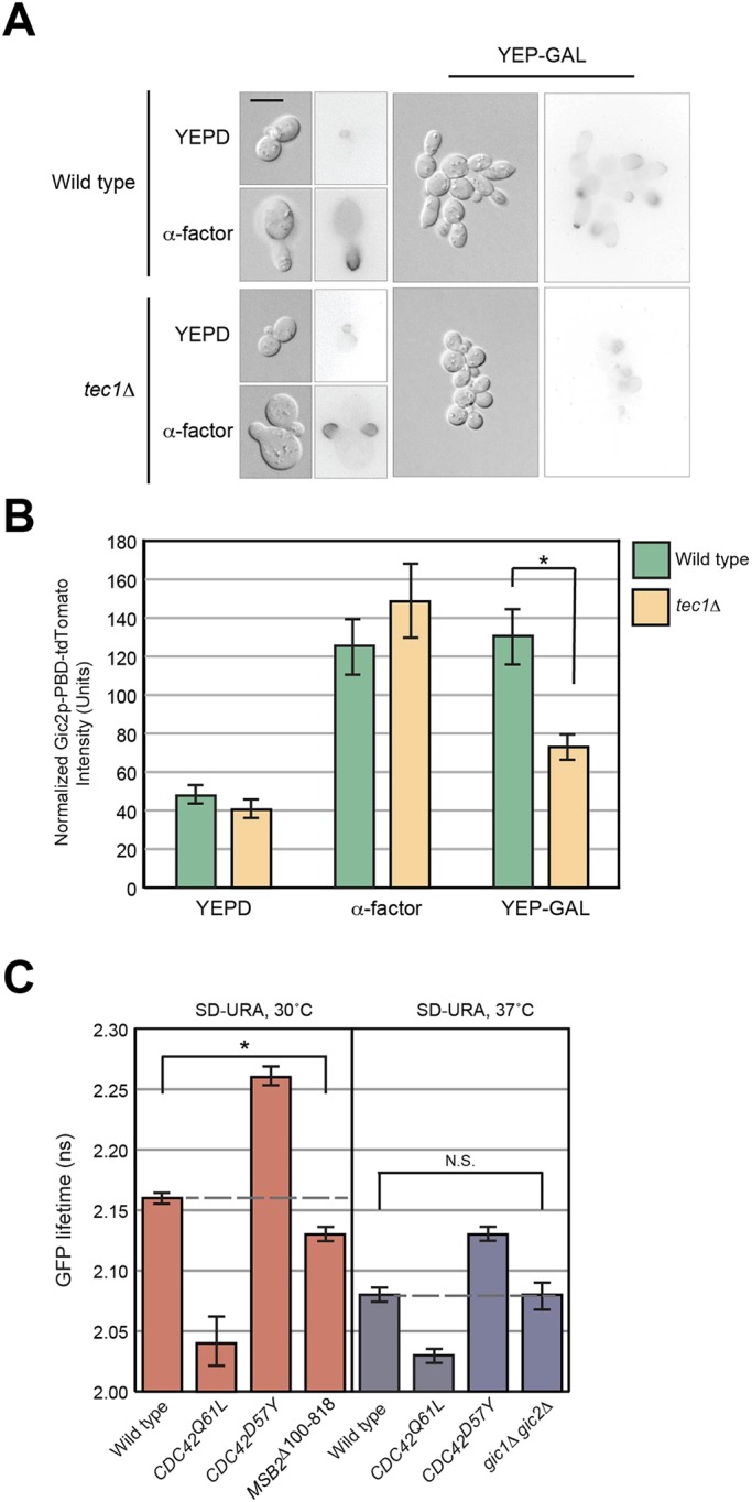
    1. Adhikari H., Vadaie N., Chow J., Caccamise L. M., Chavel C. A., Li B., Bowitch A., Stefan C. J. and Cullen P. J. (2015). Role of the unfolded protein response in regulating the mucin-dependent filamentous-growth mitogen-activated protein kinase pathway. Mol. Cell. Biol. 35, 1414-1432. 10.1128/MCB.01501-14 - DOI - PMC - PubMed
    1. Arkowitz R. A. and Iglesias P. A. (2008). Basic principles of polarity establishment and maintenance. Conference on Mechanisms of Cell Polarity. EMBO Rep. 9, 847-852. 10.1038/embor.2008.164 - DOI - PMC - PubMed
    1. Asakura T., Sasaki T., Nagano F., Satoh A., Obaishi H., Nishioka H., Imamura H., Hotta K., Tanaka K., Nakanishi H. et al. (1998). Isolation and characterization of a novel actin filament-binding protein from Saccharomyces cerevisiae. Oncogene 16, 121-130. 10.1038/sj.onc.1201487 - DOI - PubMed
    1. Ashburner M., Ball C. A., Blake J. A., Botstein D., Butler H., Cherry J. M., Davis A. P., Dolinski K., Dwight S. S., Eppig J. T. et al. (2000). Gene ontology: tool for the unification of biology. The Gene Ontology Consortium. Nat. Genet. 25, 25-29. 10.1038/75556 - DOI - PMC - PubMed

Publication types

MeSH terms

Substances