. 2020 Mar 30;133(6):jcs242412.
doi: 10.1242/jcs.242412.

PTPN23 binds the dynein adaptor BICD1 and is required for endocytic sorting of neurotrophin receptors

Affiliations

PTPN23 binds the dynein adaptor BICD1 and is required for endocytic sorting of neurotrophin receptors

Marta I Budzinska et al. J Cell Sci. .

Abstract

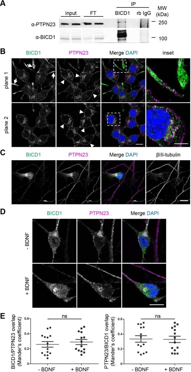
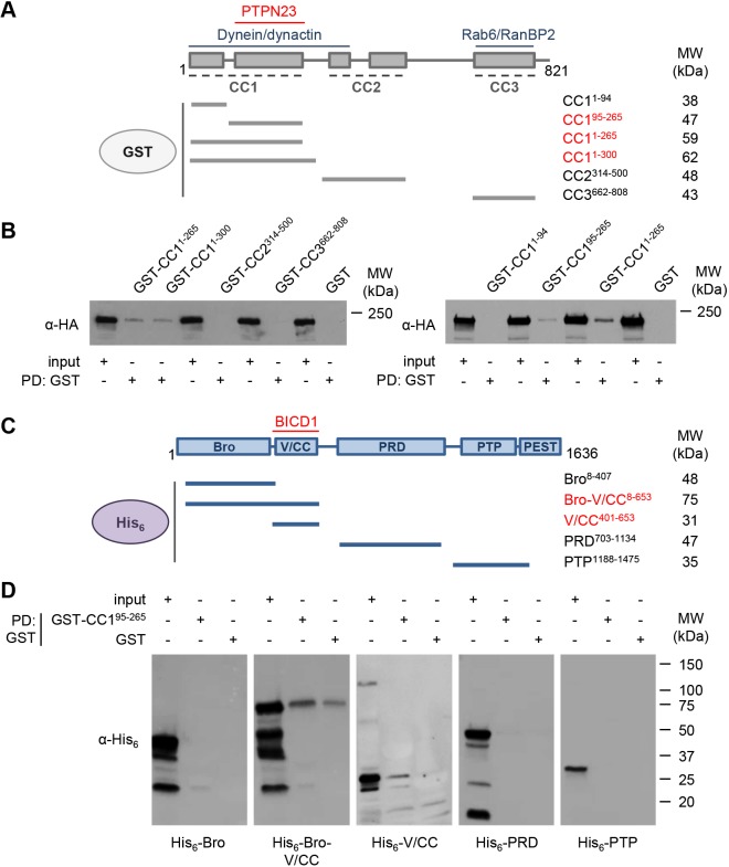
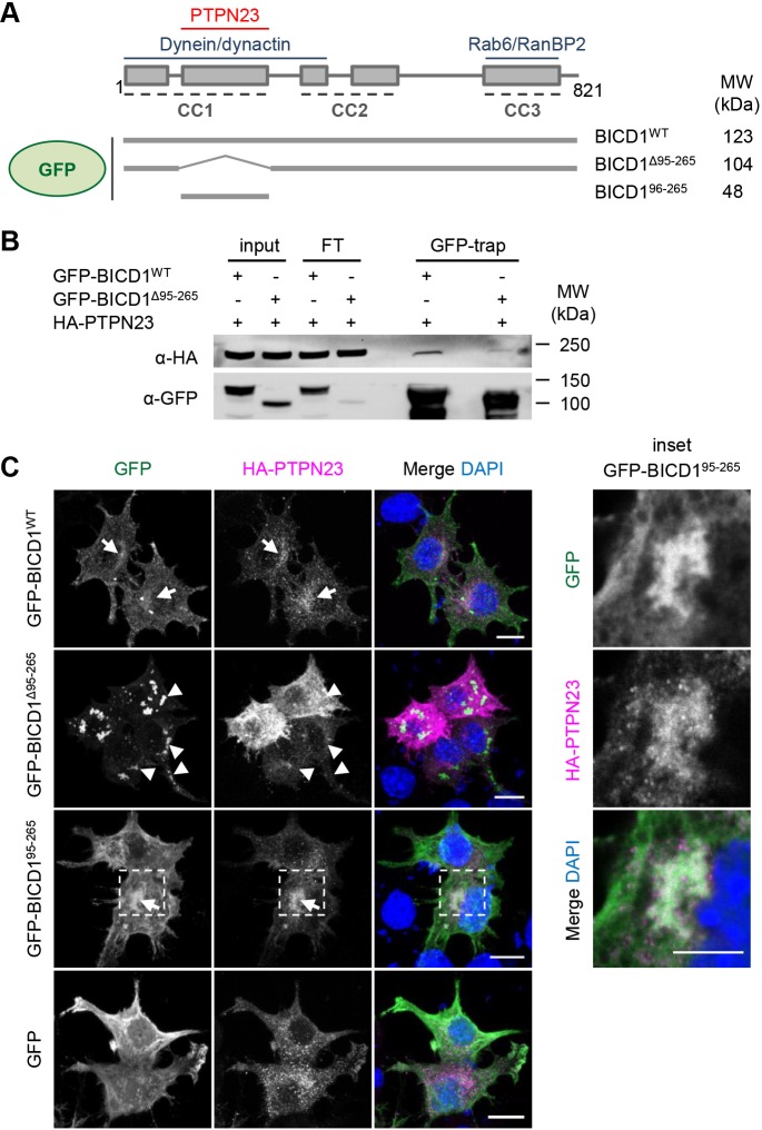
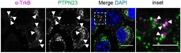
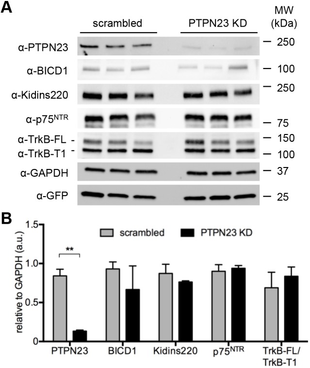
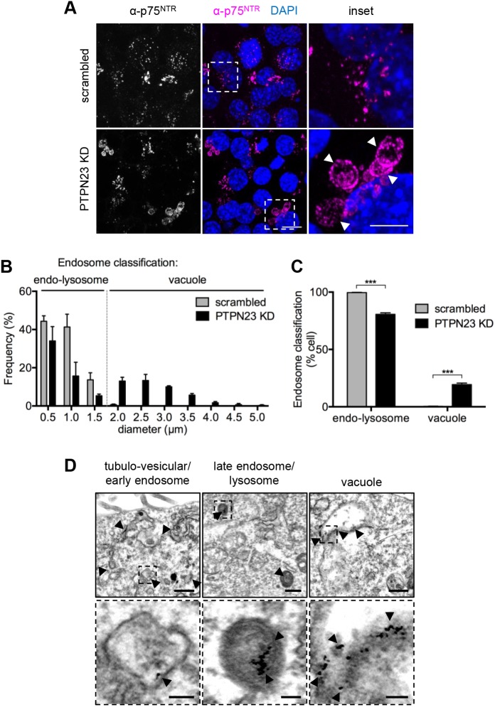
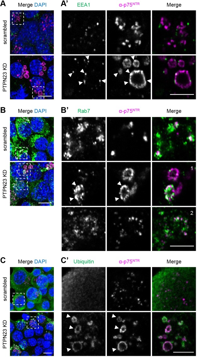
Signalling by target-derived neurotrophins is essential for the correct development of the nervous system and its maintenance throughout life. Several aspects concerning the lifecycle of neurotrophins and their receptors have been characterised over the years, including the formation, endocytosis and trafficking of signalling-competent ligand-receptor complexes. However, the molecular mechanisms directing the sorting of activated neurotrophin receptors are still elusive. Previously, our laboratory identified Bicaudal-D1 (BICD1), a dynein motor adaptor, as a key factor for lysosomal degradation of brain-derived neurotrophic factor (BDNF)-activated TrkB (also known as NTRK2) and p75NTR (also known as NGFR) in motor neurons. Here, using a proteomics approach, we identified protein tyrosine phosphatase, non-receptor type 23 (PTPN23), a member of the endosomal sorting complexes required for transport (ESCRT) machinery, in the BICD1 interactome. Molecular mapping revealed that PTPN23 is not a canonical BICD1 cargo; instead, PTPN23 binds the N-terminus of BICD1, which is also essential for the recruitment of cytoplasmic dynein. In line with the BICD1-knockdown phenotype, loss of PTPN23 leads to increased accumulation of BDNF-activated p75NTR and TrkB in swollen vacuole-like compartments, suggesting that neuronal PTPN23 is a novel regulator of the endocytic sorting of neurotrophin receptors.

Keywords: Intracellular sorting; Motor neuron; Trafficking; TrkB; p75NTR.


Conflict of interest statement

Competing interestsThe authors declare no competing or financial interests.

Figures

Fig. 1.
Fig. 1.
PTPN23 co-immunoprecipitates and colocalises with BICD1 in motor neurons and in a neuronal cell line. (A) N2A-FLAG-TrkB cell lysates were subjected to co-immunoprecipitation using Protein-G magnetic beads, pre-coated with affinity-purified anti-BICD1 polyclonal antibody or rabbit IgG as a control. Western blotting of the immunoprecipitates using anti-PTPN23 and anti-BICD1 antibodies shows specific co-immunoprecipitation of endogenous PTPN23 with BICD1 (n=5). (B) Confocal images of N2A-FLAG-TrkB cells, fixed and immunostained using anti-BICD1 and anti-PTPN23 antibodies. BICD1 is enriched in cell protrusions (arrows), whilst PTPN23 is enriched near the cell surface (arrowheads). BICD1 and PTPN23 are highly abundant in the perinuclear region (inset), where their immunostaining partially overlaps (n=6). Scale bar: 10 μm; inset: 5 μm. (C) Confocal images of ES-MNs, fixed and immunostained for BICD1, PTPN23 and the neuronal marker βIII-tubulin, showing partially overlapping and perinuclear enrichment of BICD1 and PTPN23 (n=3). Scale bar: 10 μm. (D) Confocal images of ES-MNs. Cells were starved and stimulated with or without BDNF for 1 h, fixed and immunostained for BICD1 and PTPN23 (n=2). Scale bar: 10 μm. (E) Quantification of BICD1 and PTPN23 colocalisation (from D), using Mander's coefficient. Analysis of 14 neurons per condition; P>0.05, Student's t-test (ns, not significant).
Fig. 2.
Fig. 2.
BICD1 and PTPN23 directly interact in vitro. (A) Schematic representation of BICD1 domain architecture, depicting its coiled coil domains (CC1–CC3) and highlighting the known binding regions of dynein, dynactin, Rab6 and RanBP2. Below, a schematic of GST-BICD1 fragments, with predicted molecular weights (MW; kDa). GST-BICD1 proteins in red bind HA-PTPN23 in GST pull downs. (B) GSH resin pre-loaded with GST recombinant proteins was incubated with N2A-FLAG-TrkB cell lysates, containing overexpressed HA-PTPN23 (n=2). Following GST pull downs, eluted proteins were assessed by immunoblotting using anti-HA antibodies. BICD1-CC195-265 is the shortest fragment precipitating HA-PTPN23 (n=6). (C) Schematic representation of PTPN23 domain architecture, and His6-PTPN23 fragments used for GST pull downs, with predicted MW. His6-PTPN23 proteins in red bind GST-BICD1. (D) GSH resin pre-loaded with GST-CC195-265 or GST was incubated with bacterial cell extracts containing His6-PTPN23 fragments (n=3). Pulled down proteins were eluted and assessed by immunoblotting using anti-His6 antibodies.
Fig. 3.
Fig. 3.
GFP-BICD1Δ95-265 displays reduced association with PTPN23. (A) Schematic of GFP-BICD1 proteins used for co-immunoprecipitation and confocal microscopy. (B) N2A-FLAG-TrkB cells were transfected overnight with plasmids encoding HA-PTPN23 and GFP-BICD1WT or GFP-BICD1Δ95-265, and cell extracts subjected to co-immunoprecipitation using GFP-trap beads (n=1). Eluted proteins were assessed by immunoblotting using anti-HA and anti-GFP antibodies, revealing reduced binding between mutant GFP-BICD1Δ95-265 and HA-PTPN23, relative to GFP-BICD1WT. (C) N2A-FLAG-TrkB cells were transfected overnight with plasmids expressing HA-PTPN23 and GFP-BICD1 variants or GFP as a control, fixed and immunostained using anti-HA antibody (n=3). GFP-BICD1WT and GFP-BICD195-265 (see inset) colocalise with HA-PTPN23 (arrows), whilst the aggregation-prone GFP-BICD1Δ95-265 translocates to the cell periphery (arrowheads). Images show maximum intensity Z-stack projections, acquired at 0.5 μm spacing; insets show a representative frame selected from the respective Z-stack. Scale bars: 10 μm, inset: 5 μm.
Fig. 4.
Fig. 4.
PTPN23 associates with TrkB-containing endocytic vesicles. Confocal images of anti-TrkB antibody accumulation assay in N2A-FLAG-TrkB cells. Cells were incubated for 30 min with antibody and chased for 30 min with 100 ng/ml BDNF. Following acid wash and fixation, cells were immunostained using anti-PTPN23 antibodies and Alexa Fluor 488-conjugated secondary antibodies. Alexa Fluor 555-conjugated secondary antibodies were used to reveal the localisation of TrkB (n=1). Scale bar: 10 μm; inset: 5 μm.
Fig. 5.
Fig. 5.
PTPN23 is not essential to maintain steady-state levels of NTRs. (A) N2A-FLAG-TrkB cells were transduced for 72 h with scrambled or PTPN23-targeting (PTPN23-KD) shRNAs. Cell lysates were immunoblotted for PTPN23 and NTR and related proteins of interest. GAPDH was used as a loading control, and the levels of GFP lentiviral reporter were assessed (n=3). (B) Densitometric analysis of protein levels from A, normalised to GAPDH (n=3). **P<0.01, unpaired Student's t-test.
Fig. 6.
Fig. 6.
Silencing of PTPN23 leads to p75NTR accumulation in vacuole-like compartments. (A) Confocal images of α-p75NTR accumulation assay in scrambled and PTPN23 shRNA treated N2A-FLAG-TrkB cells (n=3). Cells were incubated for 30 min with anti-p75NTR antibody and chased for 30 min with 100 ng/ml BDNF. Following acid wash and fixation, receptor localisation was revealed using Alexa Fluor 555-conjugated secondary antibody. Arrowheads indicate enlarged endocytic compartments. Images show maximum intensity Z-stack projections, acquired at 0.5 μm spacing. Scale bar: 10 μm; inset: 5 μm. (B) Diameters of 100 endosomes (per n/condition) were measured and grouped using 0.5 μm binning, in three independent experiments. Endosomes were classified as endo-lysosomes (<1.5 μm) or vacuoles (>1.5 μm). ****P<0.0001, χ2 (two-tailed chi-square) test for trend (n=3). (C) Quantification of cells containing endo-lysosomes or vacuole-like compartments (n=3); cells lacking α-p75NTR accumulation (∼50% of cells) were counted but excluded from the analysis. ***P<0.001, unpaired Student's t-test. (D) Transmission electron microscopy images showing three representative classes of organelles containing accumulated α-p75NTR-gold (arrowheads). Early and late endocytic compartments were indistinguishable between scrambled and PTPN23-KD cells, and representative images taken from PTPN23-KD and scrambled cells, respectively; vacuoles were observed only in PTPN23-KD cells. Scale bars: 500 nm; insets: 100 nm.
Fig. 7.
Fig. 7.
Vacuolar compartments, containing α-p75NTR, are sorting endosomes enriched in ubiquitylated proteins. Confocal images of α-p75NTR accumulation in scrambled and PTPN23 shRNA-treated N2A-FLAG-TrkB cells. Following acid wash and fixation, cells were immunostained using anti-EEA1 (A), anti-Rab7 (B) and anti-ubiquitin (C) antibodies (n=3). Arrowheads indicate co-localization of these proteins within enlarged endocytic compartments. (A,B,C) Merged images showing maximum intensity Z-stack projections, acquired at 0.5 μm spacing. (A′,B′,C′) Insets show representative frames selected from corresponding Z-stacks in A,B,C, respectively. Scale bars: 10 μm; insets: 5 μm.

Similar articles

Cited by

References

    1. Alazami A. M., Patel N., Shamseldin H. E., Anazi S., Al-Dosari M. S., Alzahrani F., Hijazi H., Alshammari M., Aldahmesh M. A., Salih M. A. et al. (2015). Accelerating novel candidate gene discovery in neurogenetic disorders via whole-exome sequencing of prescreened multiplex consanguineous families. Cell Rep. 10, 148-161. 10.1016/j.celrep.2014.12.015 - DOI - PubMed
    1. Ali N., Zhang L., Taylor S., Mironov A., Urbé S. and Woodman P. (2013). Recruitment of UBPY and ESCRT exchange drive HD-PTP-dependent sorting of EGFR to the MVB. Curr. Biol. 23, 453-461. 10.1016/j.cub.2013.02.033 - DOI - PubMed
    1. Ascano M., Bodmer D. and Kuruvilla R. (2012). Endocytic trafficking of neurotrophins in neural development. Trends Cell Biol. 22, 266-273. 10.1016/j.tcb.2012.02.005 - DOI - PMC - PubMed
    1. Barford K., Deppmann C. and Winckler B. (2017). The neurotrophin receptor signaling endosome: where trafficking meets signaling. Dev. Neurobiol. 77, 405-418. 10.1002/dneu.22427 - DOI - PMC - PubMed
    1. Bianco A., Dienstbier M., Salter H. K., Gatto G. and Bullock S. L. (2010). Bicaudal-D regulates fragile X mental retardation protein levels, motility, and function during neuronal morphogenesis. Curr. Biol. 20, 1487-1492. 10.1016/j.cub.2010.07.016 - DOI - PMC - PubMed

Publication types

MeSH terms

Substances