. 2020 Feb 21;367(6480):888-892.
doi: 10.1126/science.aay9813.

Angiotensin and biased analogs induce structurally distinct active conformations within a GPCR

Affiliations

Angiotensin and biased analogs induce structurally distinct active conformations within a GPCR

Laura M Wingler et al. Science. .

Abstract

Biased agonists of G protein-coupled receptors (GPCRs) preferentially activate a subset of downstream signaling pathways. In this work, we present crystal structures of angiotensin II type 1 receptor (AT1R) (2.7 to 2.9 angstroms) bound to three ligands with divergent bias profiles: the balanced endogenous agonist angiotensin II (AngII) and two strongly β-arrestin-biased analogs. Compared with other ligands, AngII promotes more-substantial rearrangements not only at the bottom of the ligand-binding pocket but also in a key polar network in the receptor core, which forms a sodium-binding site in most GPCRs. Divergences from the family consensus in this region, which appears to act as a biased signaling switch, may predispose the AT1R and certain other GPCRs (such as chemokine receptors) to adopt conformations that are capable of activating β-arrestin but not heterotrimeric Gq protein signaling.


Figures

Fig. 1.
Fig. 1.. Endogenous and biased AT1R ligands.
(A) Peptide ligands crystallized with the AT1R. Bold residues indicate mutations relative to AngII, the endogenous agonist. NMe, N-methyl. (B) Ligand activation of Gq-mediated inositol monophosphate (IP) increases and β-arrestin2 endocytosis. (C) Bias factors of ligands relative to AngII determined from the data shown in panel (B) as described in Materials and Methods. A bias factor of 1 represents a 10-fold difference in the ligand’s ability to activate the β-arrestin pathway compared to the Gq pathway. (D) Nanobody AT110i1 allosterically increases binding of AngII, TRV026, and TRV023 to purified AT1R (Ki values in table S1). (D) AngII, TRV026, and TRV023 promote interaction of FLAG-AT1R and AT110-His6, the lower affinity parental clone of AT110i1, by AlphaScreen. Data are normalized to signal without ligand. In (B),(D), and (E), the means ± standard error from three independent experiments are shown. In (C), error bars represent the standard error in bias factors derived from curve fit parameters from (B).
Fig. 2.
Fig. 2.. Comparison of AngII- and β-arrestin-biased ligand-bound AT1R.
(A) Activated AT1R bound to AngII (yellow) displays characteristic TM6 and TM7 movements stabilized by TM6 conformational locks (sticks). AT1R bound to TRV023 (0.43Å RMSD for 264 Cα atoms), and TRV026 (0.42 Å RMSD for 264 Cα atoms) is nearly identical in overall conformation. (B)-(D) Similar binding modes of TRV023 (B), TRV026 (C), and AngII (D). Dashed lines indicate hydrogen bonds. (E) AngII-AT1R ligand-binding pocket colored by Cα B-factors, highlighting the mobility of F8. (F)-(H) Electron density (gray mesh, 2Fo-Fc density contoured at 1σ) of C-terminal regions of TRV023 (F), TRV026 (G), and AngII (H) and surrounding AT1R residues. Weak density for AngII F8 and Y292 (H) indicates that the residues are structurally heterogeneous within the crystal lattice, likely because they are dynamic (13).
Fig. 3.
Fig. 3.. AngII-AT1R exhibits distinct configurations of conformational locks.
(A) View of AngII-AT1R, with key residues highlighted. (B) Movement of L1123.36 accommodates the position of AngII F8. The concomitant rotation of TM3 repositions N1113.35 outside the receptor core. TRV026 binding does not have this effect. (C),(D) Change in affinity of AT1R ligands for AT1R L112A3.36 (C) and Y292A7.43 (D) versus wild-type AT1R. Losartan is a small-molecule antagonist; TRV055 is a Gq-biased agonist with a C-terminal phenylalanine (7). Error bars represent the standard error of the difference in the log Ki values determined from 3–4 independent experiments. See also fig. S6 and table S4. For (C) and (D), * indicates statistically significant difference (p < 0.05) for mutant vs. wild-type Ki values as determined by a t-test with Holm-Sidak correction for multiple comparisons. (E) Both AngII-bound and TRV026-bound structures exhibit the outward rotation of TM6 and inward rotation of TM7, rearranging conformational locks. (F) The AngII-induced rotation of N1113.35 alters the polar network at the canonical sodium binding site in the receptor core involving D742.50, N2957.46, and N2987.49 (of the NPxxY motif).
Fig. 4.
Fig. 4.. Structural diversity in the polar core of GPCRs.
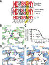
(A) Conservation of sodium coordination residues (26), with Family A GPCR consensus residues in red. Many chemokine receptors (36) deviate from the consensus sequence. (B) Sodium binding in the δ-opioid receptor. N3.35 and S7.46 are found in the sodium coordination sphere. (C) Effect of sodium on AngII binding to wild-type AT1R in membranes. Competition radioligand binding was performed in buffer containing 150 mM NaCl (log KiAngII = −7.54 ± 0.06, Ki = 29 nM; Kd[3H]-olmesartan = 1.2 ± 0.1 nM) or lacking sodium and containing 150 mM KCl (log KiAngII = −7.73 ± 0.02, Ki = 19 nM; Kd[3H]-olmesartan = 1.2 ± 0.3 nM). The means ± standard error from three independent experiments are shown. (D),(E) Residues associated with sodium binding make up the AT1R polar network. (D) The N1113.35- N2957.46 hydrogen bond stabilizes the inactive state in antagonist (ZD7155)-bound AT1R. Upon TRV026 binding, TM7 movement disrupts the hydrogen bond. (E) AngII binding rotates N1113.35 away from the polar core, yielding a second active conformation.

References

    1. Lefkowitz RJ, A brief history of G-protein coupled receptors (Nobel Lecture). Angew. Chem. Int. Ed. Engl. 52, 6366–6378 (2013). - PubMed
    1. Peterson YK, Luttrell LM, The diverse roles of arrestin scaffolds in G protein-coupled receptor signaling. Pharmacol. Rev. 69, 256–297 (2017). - PMC - PubMed
    1. Rankovic Z, Brust TF, Bohn LM, Biased agonism: An emerging paradigm in GPCR drug discovery. Bioorg. Med. Chem. Lett. 26, 241–250 (2016). - PMC - PubMed
    1. Holloway AC et al., Side-chain substitutions within angiotensin II reveal different requirements for signaling, internalization, and phosphorylation of type 1A angiotensin receptors. Mol. Pharmacol. 61, 768–777 (2002). - PubMed
    1. Wei H et al., Independent beta-arrestin 2 and G protein-mediated pathways for angiotensin II activation of extracellular signal-regulated kinases 1 and 2. Proc. Natl. Acad. Sci. USA 100, 10782–10787 (2003). - PMC - PubMed

Publication types