

Can diffusion weighted imaging be used as an alternative to contrast-enhanced imaging on magnetic resonance enterography for the assessment of active inflammation in Crohn disease?
- PMID: 32080107
- PMCID: PMC7034637
- DOI: 10.1097/MD.0000000000019202
Can diffusion weighted imaging be used as an alternative to contrast-enhanced imaging on magnetic resonance enterography for the assessment of active inflammation in Crohn disease?
Abstract
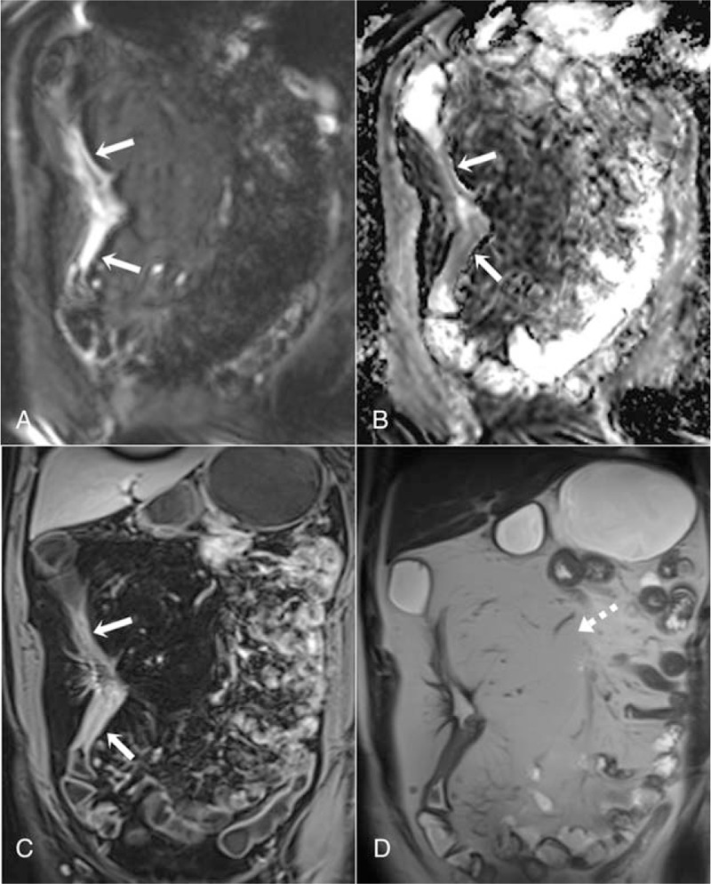
The present study aimed to investigate the potential use of T2-weighted sequences with diffusion weighted imaging (DWI) in magnetic resonance (MR) enterography instead of conventional contrast-enhanced MR imaging (MRI) sequences for the evaluation of active inflammation in Crohn disease.Two-hundred thirteen intestinal segments of 43 patients, who underwent colonoscopy within 2 weeks before or after MR enterography were evaluated in this retrospective study. DWI sequences, T2-weighted sequences, and contrast-enhanced T1-weighted sequences were acquired in the MR enterography scan after cleaning of the bowel and using an oral contrast agent. First, the intestinal segments that had active inflammation in MR enterography were qualitatively evaluated in T2-weighted and contrast-enhanced T1-weighted sequences and then MR activity index (MRAI 1) and MRAI 2 were formed with and without contrast-enhanced sequences in 2 separate sessions.The correlation coefficient between contrast enhanced and DWI MR enterography scores (MRAI 1 and MRAI 2) of intestinal inflammation was 0.97 for all segments. In addition, separate correlation coefficients were calculated for terminal ileum, right colon, transverse colon, left colon, and rectum, and there was a strong correlation between the MRAI 1 and MRAI 2 scores of each segment (r = 0.86-0.97, P < .001). On the other hand, MR enterography had 88.7% sensitivity, 97.9% specificity, 95.5% positive predictive value, 94.6% negative predictive value, and 94.8% accuracy for detection of active inflammation in all intestinal segments in Crohn disease.DWI and T2-weighted sequences acquired with cleaning of the bowel can be used instead of contrast-enhanced MRI sequences for the evaluation of active inflammation in Crohn disease.
Conflict of interest statement
The authors have no conflicts of interest to disclose.
Figures


References
-
- Scaldaferri F, Fiocchi C. Inflammatory bowel disease: progress and current concepts of etiopathogenesis. J Dig Dis 2007;8:171–8. - PubMed
-
- Fedorak RN. Thompson ABR, Shaffer EA. Inflammatory bowel disease. AstraZeneca Canada, First principles of gastroenterology. Toronto, Canada: 2008.
-
- Rondonotti E, Spada C, Adler S, et al. Small-bowel capsule endoscopy and device-assisted enteroscopy for diagnosis and treatment of small-bowel disorders: European Society of Gastrointestinal Endoscopy (ESGE) Technical Review. Endoscopy 2018;50:423–46. - PubMed
-
- Barber JL, Shah N, Watson TA. Early onset inflammatory bowel disease–What the radiologist needs to know. Eur J Radiol 2018;106:173–82. - PubMed
MeSH terms
Substances
LinkOut - more resources
Full Text Sources
Medical
