Comparative Study
. 2020 Feb 20;10(1):3036.
doi: 10.1038/s41598-020-59924-1.

Comparative in-silico proteomic analysis discerns potential granuloma proteins of Yersinia pseudotuberculosis

Affiliations
Comparative Study

Comparative in-silico proteomic analysis discerns potential granuloma proteins of Yersinia pseudotuberculosis

Manisha Aswal et al. Sci Rep. .

Abstract

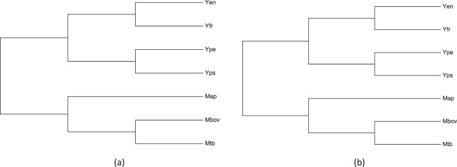
Yersinia pseudotuberculosis is one of the three pathogenic species of the genus Yersinia. Most studies regarding pathogenesis of Y. pseudotuberculosis are based on the proteins related to Type III secretion system, which is a well-known primary virulence factor in pathogenic Gram-negative bacteria, including Y. pseudotuberculosis. Information related to the factors involved in Y. pseudotuberculosis granuloma formation is scarce. In the present study we have used a computational approach to identify proteins that might be potentially involved in formation of Y. pseudotuberculosis granuloma. A comparative proteome analysis and conserved orthologous protein identification was performed between two different genera of bacteria - Mycobacterium and Yersinia, their only common pathogenic trait being ability to form necrotizing granuloma. Comprehensive analysis of orthologous proteins was performed in proteomes of seven bacterial species. This included M. tuberculosis, M. bovis and M. avium paratuberculosis - the known granuloma forming Mycobacterium species, Y. pestis and Y. frederiksenii - the non-granuloma forming Yersinia species and, Y. enterocolitica - that forms micro-granuloma and, Y. pseudotuberculosis - a prominent granuloma forming Yersinia species. In silico proteome analysis indicated that seven proteins (UniProt id A0A0U1QT64, A0A0U1QTE0, A0A0U1QWK3, A0A0U1R1R0, A0A0U1R1Z2, A0A0U1R2S7, A7FMD4) might play some role in Y. pseudotuberculosis granuloma. Validation of the probable involvement of the seven proposed Y. pseudotuberculosis granuloma proteins was done using transcriptome data analysis and, by mapping on a composite protein-protein interaction map of experimentally proved M. tuberculosis granuloma proteins (RD1 locus proteins, ESAT-6 secretion system proteins and intra-macrophage secreted proteins). Though, additional experiments involving knocking out of each of these seven proteins are required to confirm their role in Y. pseudotuberculosis granuloma our study can serve as a basis for further studies on Y. pseudotuberculosis granuloma.


Conflict of interest statement

The authors declare no competing interests.

Figures

Figure 1
Figure 1
Relatedness among the seven bacterial species on the basis of their genome and proteomes. Cladograms were generated using Neighbor-Joining method using (a) average nucleotide identity (ANI) and (b) Percentage of conserved proteins (PCOP).
Figure 2
Figure 2
Number of proteins in mutually exclusive protein clusters formed due to different combinations of seven proteomes (M. tuberculosis, M. bovis, M. paratuberculosis and non-granuloma forming Yersinia species like Y. pseudotuberculosis, Y. enterocolitica and Y. frederiksenii). Solid dots show presence of proteins in the corresponding proteome and line between two solid dots shows presence of orthologous proteins in the two proteomes. The vertical bars show the number of shared homologous proteins (orthologous proteins) among the proteomes. Single solid dot represents species unique proteins. The plot was drawn from UpSet plot tool (https://gehlenborglab.shinyapps.io/upsetr/). Panel (a–c) Shows combination of proteomes which shared >10, ≤10 and 0 orthologous proteins respectively. ‘*’ represent proteins shared among all seven proteomes, ‘$’ and ‘#’ shows intra-genus conserved proteins of Mycobacterium spp. and Yersinia spp respectively.
Figure 3
Figure 3
Functional enrichment analysis of different mutually exclusive protein clusters. The enrichment analysis was done for six sets, Set I - inter-genus, Set II - intra-genus: (A) Mycobacterium spp. and (B) Yersinia spp., and Mycobacterium spp. with Ype Set III; Yen Set IV; Yps Set V and Yfr Set VI. Top 10 enriched GO terms of GO Biological Processes (BP), GO Cellular Components (CC), and GO Molecular Functions (MF) are shown in panels a–c, respectively. Red color shows absence of function where as white shows presence.
Figure 4
Figure 4
A composite protein-protein interaction network map of Mtb RD1 locus proteins, ESAT-6 secretion system proteins and intra-macrophage secreted proteins, constructed using STRING database.The Mtb orthologs of the seven proposed Yps proteins (Rv0366c, Rv0712, Rv1458c, Rv2858c (aldc), Rv3096, Rv3173c, and Rv3382c (lytB2)) are marked in red colored rectangular boxes. The colour and thickness of edges (lines connecting two proteins or nodes) indicates type and confidence of interaction, respectively. The color coding of edges are as follows: Red line - indicates the presence of fusion evidence; Green line - neighborhood evidence; Blue line - cooccurrence evidence; Purple line - experimental evidence; Yellow line - textmining evidence; Light blue line - database evidence; Black line - coexpression evidence.

References

    1. Long C, et al. Yersinia pseudotuberculosis and Y. enterocolitica infections, FoodNet, 1996–2007. Emerg. Infect. Dis. 2010;16:566–567. doi: 10.3201/eid1603.091106. - DOI - PMC - PubMed
    1. Pujol C, Bliska JB. The ability to replicate in macrophages is conserved between Yersinia pestis and Yersinia pseudotuberculosis. Infect. Immun. 2003;71:5892–5899. doi: 10.1128/IAI.71.10.5892-5899.2003. - DOI - PMC - PubMed
    1. McNally A, Thomson NR, Reuter S, Wren BW. Add, stir and reduce: Yersinia spp. as model bacteria for pathogen evolution. Nat. Rev. Microbiol. 2016;14:177–190. doi: 10.1038/nrmicro.2015.29. - DOI - PubMed
    1. Reuter S, et al. Parallel independent evolution of pathogenicity within the genus Yersinia. Proc. Natl Acad. Sci. USA. 2014;111:6768–6773. doi: 10.1073/pnas.1317161111. - DOI - PMC - PubMed
    1. Westermark L, Fahlgren A, Fallman M. Yersinia pseudotuberculosis efficiently escapes polymorphonuclear neutrophils during early infection. Infect. Immun. 2014;82:1181–1191. doi: 10.1128/IAI.01634-13. - DOI - PMC - PubMed

Publication types

LinkOut - more resources