. 2020 Feb 20;10(1):3007.
doi: 10.1038/s41598-020-59940-1.

Keel bone fractures induce a depressive-like state in laying hens

Affiliations

Keel bone fractures induce a depressive-like state in laying hens

E A Armstrong et al. Sci Rep. .

Abstract

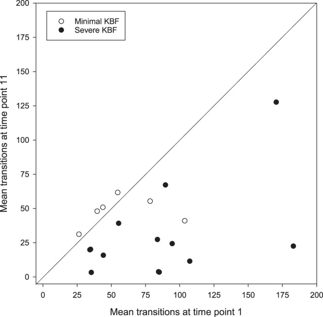
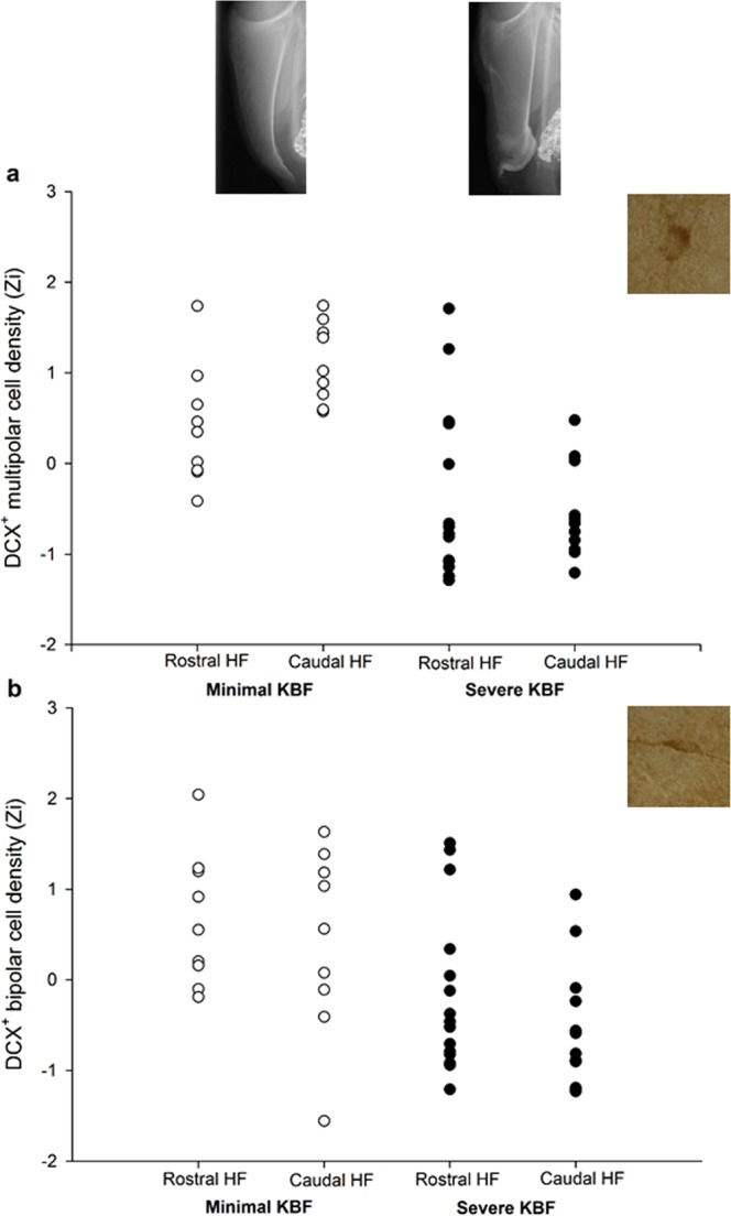
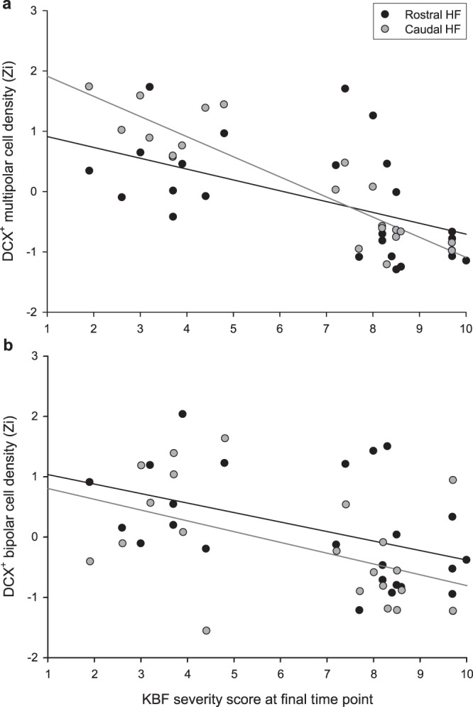
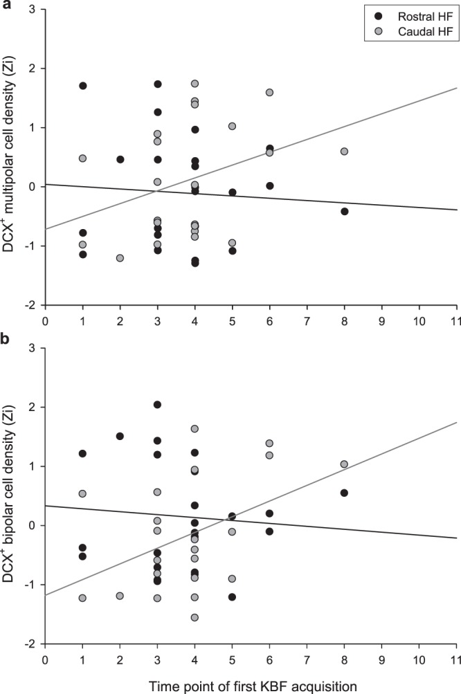
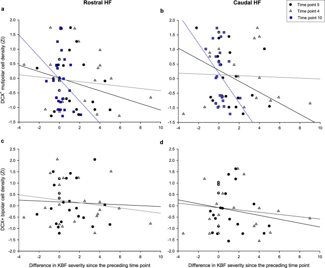
In commercial flocks of laying hens, keel bone fractures (KBFs) are prevalent and associated with behavioural indicators of pain. However, whether their impact is severe enough to induce a depressive-like state of chronic stress is unknown. As chronic stress downregulates adult hippocampal neurogenesis (AHN) in mammals and birds, we employ this measure as a neural biomarker of subjective welfare state. Radiographs obtained longitudinally from Lohmann Brown laying hens housed in a commercial multi-tier aviary were used to score the severity of naturally-occurring KBFs between the ages of 21-62 weeks. Individual birds' transitions between aviary zones were also recorded. Focal hens with severe KBFs at 3-4 weeks prior to sampling (n = 15) had lower densities of immature doublecortin-positive (DCX+) multipolar and bipolar neurons in the hippocampal formation than focal hens with minimal fractures (n = 9). KBF severity scores at this time also negatively predicted DCX+ cell numbers on an individual level, while hens that acquired fractures earlier in their lives had fewer DCX+ neurons in the caudal hippocampal formation. Activity levels 3-4 weeks prior to sampling were not associated with AHN. KBFs thus lead to a negative affective state lasting at least 3-4 weeks, and management steps to reduce their occurrence are likely to have significant welfare benefits.


Conflict of interest statement

The authors declare no competing interests.

Figures

Figure 1
Figure 1
The mean number of vertical aviary transitions made by hens which had developed minimal versus severe keel bone fractures (KBFs) by time point 11, made during the 6 days preceding the first (T1) and final (T11) radiograph time points. Line of equality represents equal activity at both time points. Some data points are overlapping.
Figure 2
Figure 2
Densities of (a) multipolar and (b) bipolar DCX-stained cells in the rostral and caudal HF subregions of hens with minimal versus severe keel bone fractures (KBFs) at time point 11, normalised (Zi) for staining batch. Example radiographs reflect the mean severity scores of the minimal and severe KBF groups: 3.5 and 8.5 respectively. Representative DCX+ stained cells of both morphologies are displayed at 100X magnification.
Figure 3
Figure 3
Relationship between keel bone fracture (KBF) score at the final time point (ranging from 0 = no KBF to 10 = extremely severe) and density of DCX-positive (a) multipolar and (b) bipolar cells in the rostral and caudal HF subregions, normalised (Zi) for staining batch. Simple linear regression lines are plotted for cell densities in the rostral (black) and caudal (grey) HF subregions.
Figure 4
Figure 4
Relationship between the time point at which individual hens first developed a keel bone fracture (KBF) and their density of DCX-positive (a) multipolar and (b) bipolar cells in the rostral and caudal HF, normalised (Zi) for staining batch. Simple linear regression lines are plotted for cell densities in the rostral (black) and caudal (grey) HF subregions.
Figure 5
Figure 5
The relationship between the difference in keel bone fracture (KBF) severity score since the preceding time point and densities of DCX+ (a) rostral multipolar, (b) caudal multipolar, (c) rostral bipolar and (d) caudal bipolar cells, for time points wherein this change in severity predicted cell densities at end of life. Simple linear regression lines are plotted for time points 5 (black), 4 (grey) and 10 (blue).

References

    1. Heerkens, J. L. T. et al. In 9th European Poultry Conference. (eds. R. Tauson, H. J. Blokhuis, L. Berg, & A. Elson) (2013).
    1. Petrik MT, Geurin MT, Widowski TM. On-farm comparison of keel fractureprevalence and other welfare indicators in conventional cage and floor-housed laying hens in Ontario, Canada. Poultry Science. 2015;94:579–585. doi: 10.3382/ps/pev039. - DOI - PubMed
    1. Riber AB, Hinrichsen LK. Keel-bone damage and foot injuries in commercial laying hens in Denmark. Animal Welfare. 2016;25:179–184. doi: 10.7120/09627286.25.2.179. - DOI
    1. Rodenburg TB, et al. Welfare assessment of laying hens in furnished cages and non-cage systems: an on-farm comparison. Animal Welfare. 2008;17:363–373.
    1. Kappeli S, Gebhardt-Henrich SG, Frohlich E, Pfulg A, Stoffel MH. Prevalence of keel bone deformities in Swiss laying hens. British Poultry Science. 2011;52:531–536. doi: 10.1080/00071668.2011.615059. - DOI - PubMed

Publication types

MeSH terms