. 2020 Feb 20;10(1):3095.
doi: 10.1038/s41598-020-59868-6.

Cardiac injury modulates critical components of prostaglandin E2 signaling during zebrafish heart regeneration

Affiliations

Cardiac injury modulates critical components of prostaglandin E2 signaling during zebrafish heart regeneration

MaryLynn FitzSimons et al. Sci Rep. .

Abstract

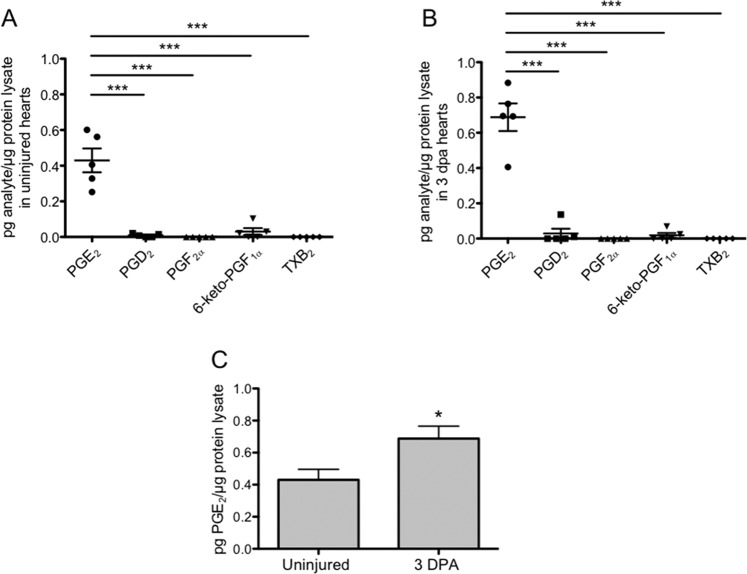
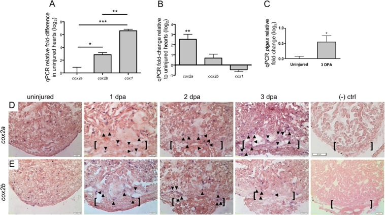
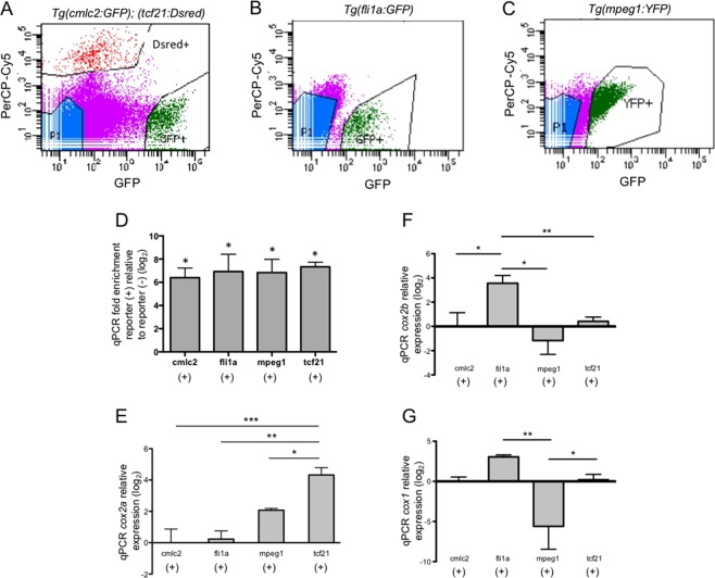
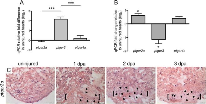
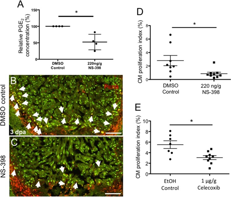
The inability to effectively stimulate cardiomyocyte proliferation remains a principle barrier to regeneration in the adult human heart. A tightly regulated, acute inflammatory response mediated by a range of cell types is required to initiate regenerative processes. Prostaglandin E2 (PGE2), a potent lipid signaling molecule induced by inflammation, has been shown to promote regeneration and cell proliferation; however, the dynamics of PGE2 signaling in the context of heart regeneration remain underexplored. Here, we employ the regeneration-competent zebrafish to characterize components of the PGE2 signaling circuit following cardiac injury. In the regenerating adult heart, we documented an increase in PGE2 levels, concurrent with upregulation of cox2a and ptges, two genes critical for PGE2 synthesis. Furthermore, we identified the epicardium as the most prominent site for cox2a expression, thereby suggesting a role for this tissue as an inflammatory mediator. Injury also drove the opposing expression of PGE2 receptors, upregulating pro-restorative ptger2a and downregulating the opposing receptor ptger3. Importantly, treatment with pharmacological inhibitors of Cox2 activity suppressed both production of PGE2, and the proliferation of cardiomyocytes. These results suggest that injury-induced PGE2 signaling is key to stimulating cardiomyocyte proliferation during regeneration.


Conflict of interest statement

The authors declare no competing interests.

Figures

Figure 1
Figure 1
Cardiac injury triggers an elevation in PGE2 synthesis. (A,B) LC-MS/MS profiling showed that PGE2 concentrations were significantly elevated above all other prostaglandin species analyzed in both the uninjured heart (A), and at 3 dpa (B). (mean ± s.e.m. n = 5 biological replicates; 5 pooled ventricles per replicate. One-way ANOVA followed by Tukey’s multiple comparisons test. ***P < 0.001). (C) LC-MS/MS analysis showed that PGE2 concentrations were significantly higher at 3 dpa, relative to uninjured hearts. (mean ± s.e.m. n = 5 biological replicates; 5 pooled ventricles per replicate. Student’s t-test. *P < 0.05).
Figure 2
Figure 2
Enzymes critical to PGE2 production are upregulated in regenerating adult hearts. (A) qPCR studies demonstrated that cox1 levels in uninjured zebrafish ventricles were significantly higher than either cox2a or cox2b transcripts. Gene expression was calculated relative to cox2a (mean ± s.e.m. n = 4–5 biological replicates; 3–5 pooled ventricles per replicate. One-way ANOVA followed by Tukey’s multiple comparisons test. *P < 0.05, **P < 0.01, ***P < 0.001). (B) At 3 dpa, cox2a is the only Cox isozyme significantly upregulated relative to uninjured hearts, as determined by qPCR. (mean ± s.e.m. n = 4–5 biological replicates; 3–5 pooled ventricles per replicate. Student’s t-test. **P < 0.01). (C) Relative to uninjured hearts, ptges is significantly upregulated at 3 dpa. (mean ± s.e.m. n = 4 biological replicates; 3–5 pooled ventricles per replicate. Student’s t-test. *P < 0.05). (D,E) Representative images of cox2a (D) and cox2b (E) expression domains as revealed by in situ hybridization studies. (n = 4 biological replicates; brackets = approximate amputation zone; arrowheads demarcate signal within the injury zone). Representative (−) control 2 dpa hearts were hybridized with cox2a riboprobe without anti-DIG antibody (top right panel) or with anti-DIG only (bottom right panel). (n = 2 biological replicates).
Figure 3
Figure 3
Cox2a expression is highest in epicardial cells at 3 dpa. (A–C) Representative FACS dot plots displaying gating parameters used to isolate cells from (A) Tg(cmlc2:EGFP); Tg(tcf21:DsRed), (B) Tg(fli1a:eGFP), (C) Tg(mpeg1:YFP) reporter lines at 3 dpa. (D) qPCR analyses of population specific markers within reporter(+) and reporter(−) cells validate the purity of FACS isolated cells. (mean ± s.e.m. n = 3 biological replicate for each cell type. Student’s t-test *P < 0.05) (E) qPCR studies in 3 dpa FACS sorted cells showed cox2a expression is significantly higher in tcf21(+) cells, relative to all other cell types examined. (F) qPCR of FACS sorted cells showed that at 3 dpa, cox2b expression was significantly higher in fli1a(+) cells relative to all other cell types examined. (G) There was no significant difference in cox1 expression among the resident cardiac cells assayed. cox1 expression in mpeg1(+) cells was significantly lower than that observed in fli1a(+) and tcf21(+) cells. Gene expression was calculated relative to cmlc2(+) cells. (mean = ±s.e.m. n = 2–5 biological replicates; 12–45 pooled ventricles per replicate. One-way ANOVA followed by Tukey’s multiple comparisons test. *P < 0.05, **P < 0.01, ***P < 0.001).
Figure 4
Figure 4
Injury activates a differential shift in PGE2 receptor expression in the heart. (A) qPCR determination of PGE2 receptor expression showed that in uninjured hearts, ptger3 levels were significantly higher than ptger2a or ptger4a. Gene expression was calculated relative to ptger2a. (mean ± s.e.m. n = 4–5 biological replicates; 3–5 pooled ventricles per replicate. One-way ANOVA followed by Tukey’s multiple comparisons test. ***P < 0.001). (B) Relative to uninjured hearts, expression of ptger2a was significantly upregulated, while ptger3 was significantly downregulated at 3 dpa. (mean ± s.e.m. n = 5 biological replicates; 3–5 pooled ventricles per replicate. Student’s t-test. *P < 0.05). (C) Representative in situ hybridization images of ptger2a expression in uninjured, 1, 2 and 3 dpa hearts. (n = 4 biological replicates, brackets mark approximate amputation zone; arrowheads demarcate signal within the injury zone).
Figure 5
Figure 5
Activation of the Cox2-PGE2 circuit stimulates cardiomyocyte proliferation. Zebrafish were subjected to ventricular amputation and treated with daily intraperitoneal (IP) injections of either vehicle control, NS-398 or Celecoxib. Hearts were collected for analysis at 3 dpa. (A) Treatment with NS-398 reduced PGE2 concentrations in ventricles by ~47%, relative to DMSO controls. (mean ± s.e.m. n = 4 biological replicate for each group; 6 pooled ventricles from weight-matched clutch mates per replicate. Student’s t-test. *P < 0.05). (B,C) Representative images of 3 dpa injured hearts treated daily with a DMSO control (B) or NS-398 (C). Hearts were stained with Mef2 (green), and Pcna (red). Mef2+Pcna+ cells mark proliferating CMs, highlighted by white arrows. Scale bars represent 50 μM. (D-E) CM proliferation indices were calculated as a percentage of Mef2(+)Pcna(+) cells relative to the total number of Mef2(+) cells in a defined area adjacent to the injury. (D) CM proliferation was reduced by more than 69% in animals treated with NS-398 when compared to controls. (E) Reduction of CM proliferation was greater than 54% in animals treated daily with Celecoxib, relative to controls. (mean ± s.e.m. n = 7–9 hearts from clutchmates; three sections were quantified per heart and results averaged. Student’s t-test. *P < 0.05).

References

    1. Finegold JA, Asaria P, Francis DP. Mortality from ischaemic heart disease by country, region, and age: statistics from World Health Organisation and United Nations. Int. J. Cardiol. 2013;168:934–945. doi: 10.1016/j.ijcard.2012.10.046. - DOI - PMC - PubMed
    1. Cano-Martinez A, et al. Functional and structural regeneration in the axolotl heart (Ambystoma mexicanum) after partial ventricular amputation. Arch. Cardiol. Mex. 2010;80:79–86. - PubMed
    1. Oberpriller JO, Oberpriller JC. Response of the adult newt ventricle to injury. J. Exp. Zool. 1974;187:249–253. doi: 10.1002/jez.1401870208. - DOI - PubMed
    1. Porrello ER, et al. Transient regenerative potential of the neonatal mouse heart. Sci. 2011;331:1078–1080. doi: 10.1126/science.1200708. - DOI - PMC - PubMed
    1. Poss KD, Wilson LG, Keating MT. Heart regeneration in zebrafish. Sci. 2002;298:2188–2190. doi: 10.1126/science.1077857. - DOI - PubMed

Publication types