

Transcytosis of Bacillus subtilis extracellular vesicles through an in vitro intestinal epithelial cell model
- PMID: 32080346
- PMCID: PMC7033168
- DOI: 10.1038/s41598-020-60077-4
Transcytosis of Bacillus subtilis extracellular vesicles through an in vitro intestinal epithelial cell model
Abstract
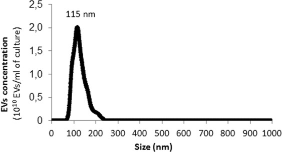
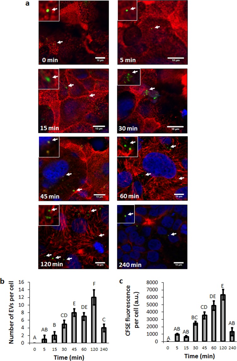
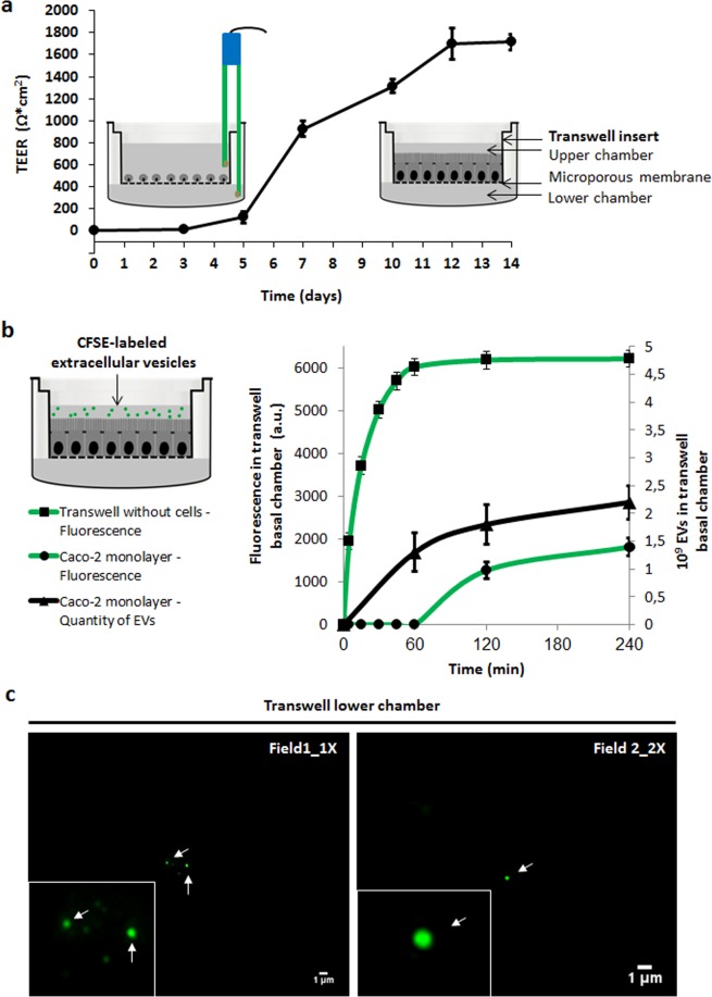
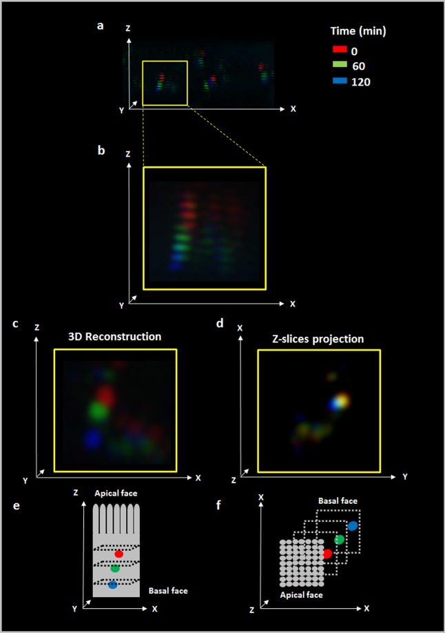
Bacterial EVs have been related to inter-kingdom communication between probiotic/pathogenic bacteria and their hosts. Our aim was to investigate the transcytosis process of B. subtilis EVs using an in vitro intestinal epithelial cell model. In this study, using Confocal Laser Scanning Microscopy, we report that uptake and internalization of CFSE-labeled B. subtilis EVs (115 nm ± 27 nm) by Caco-2 cells are time-dependent. To study the transcytosis process we used a transwell system and EVs were quantified in the lower chamber by Fluorescence and Nanoparticle Tracking Analysis measurements. Intact EVs are transported across a polarized cell monolayer at 60-120 min and increased after 240 min with an estimated average uptake efficiency of 30% and this process is dose-dependent. EVs movement into intestinal epithelial cells was mainly through Z axis and scarcely on X and Y axis. This work demonstrates that EVs could be transported across the gastrointestinal epithelium. We speculate this mechanism could be the first step allowing EVs to reach the bloodstream for further delivery up to extraintestinal tissues and organs. The expression and further encapsulation of bioactive molecules into natural nanoparticles produced by probiotic bacteria could have practical implications in food, nutraceuticals and clinical therapies.
Conflict of interest statement
The authors declare no competing interests.
Figures
Similar articles
-
Immunomodulatory properties of Bacillus subtilis extracellular vesicles on rainbow trout intestinal cells and splenic leukocytes.Front Immunol. 2024 May 7;15:1394501. doi: 10.3389/fimmu.2024.1394501. eCollection 2024. Front Immunol. 2024. PMID: 38774883 Free PMC article.
-
Polarized Secretion of Extracellular Vesicles by Mammary Epithelia.J Mammary Gland Biol Neoplasia. 2018 Sep;23(3):165-176. doi: 10.1007/s10911-018-9402-6. Epub 2018 Jul 3. J Mammary Gland Biol Neoplasia. 2018. PMID: 29968174 Free PMC article.
-
Yersinia pseudotuberculosis induces transcytosis of nanoparticles across human intestinal villus epithelium via invasin-dependent macropinocytosis.Lab Invest. 2008 Nov;88(11):1215-26. doi: 10.1038/labinvest.2008.86. Lab Invest. 2008. PMID: 18810251
-
Extracellular vesicles through the blood-brain barrier: a review.Fluids Barriers CNS. 2022 Jul 25;19(1):60. doi: 10.1186/s12987-022-00359-3. Fluids Barriers CNS. 2022. PMID: 35879759 Free PMC article. Review.
-
Cell-to-Cell Communication by Host-Released Extracellular Vesicles in the Gut: Implications in Health and Disease.Int J Mol Sci. 2021 Feb 23;22(4):2213. doi: 10.3390/ijms22042213. Int J Mol Sci. 2021. PMID: 33672304 Free PMC article. Review.
Cited by
-
Bacterial Membrane Vesicles in Pneumonia: From Mediators of Virulence to Innovative Vaccine Candidates.Int J Mol Sci. 2021 Apr 8;22(8):3858. doi: 10.3390/ijms22083858. Int J Mol Sci. 2021. PMID: 33917862 Free PMC article. Review.
-
Internalization of extracellular vesicles from Lactobacillus johnsonii N6.2 elicit an RNA sensory response in human pancreatic cell lines.J Extracell Biol. 2023 Jul;2(7):e101. doi: 10.1002/jex2.101. Epub 2023 Jul 18. J Extracell Biol. 2023. PMID: 37720361 Free PMC article.
-
Emerging role of bacterial outer membrane vesicle in gastrointestinal tract.Gut Pathog. 2023 Apr 27;15(1):20. doi: 10.1186/s13099-023-00543-2. Gut Pathog. 2023. PMID: 37106359 Free PMC article. Review.
-
Delivery of Toxins and Effectors by Bacterial Membrane Vesicles.Toxins (Basel). 2021 Nov 26;13(12):845. doi: 10.3390/toxins13120845. Toxins (Basel). 2021. PMID: 34941684 Free PMC article. Review.
-
Biocompatibility analysis of high molecular weight chitosan obtained from Pleoticus muelleri shrimps. Evaluation in prokaryotic and eukaryotic cells.Biochem Biophys Rep. 2020 Nov 12;24:100842. doi: 10.1016/j.bbrep.2020.100842. eCollection 2020 Dec. Biochem Biophys Rep. 2020. PMID: 33241127 Free PMC article.
References
-
- Hill, C. et al. STATEMENTS The International Scientific Association for. 11 (2014).
Publication types
MeSH terms
LinkOut - more resources
Full Text Sources
Other Literature Sources
