Review
. 2020 Sep;17(9):585-607.
doi: 10.1038/s41569-020-0339-2. Epub 2020 Feb 20.

Mechanisms of diabetic cardiomyopathy and potential therapeutic strategies: preclinical and clinical evidence

Affiliations
Review

Mechanisms of diabetic cardiomyopathy and potential therapeutic strategies: preclinical and clinical evidence

Yi Tan et al. Nat Rev Cardiol. 2020 Sep.

Abstract

The pathogenesis and clinical features of diabetic cardiomyopathy have been well-studied in the past decade, but effective approaches to prevent and treat this disease are limited. Diabetic cardiomyopathy occurs as a result of the dysregulated glucose and lipid metabolism associated with diabetes mellitus, which leads to increased oxidative stress and the activation of multiple inflammatory pathways that mediate cellular and extracellular injury, pathological cardiac remodelling, and diastolic and systolic dysfunction. Preclinical studies in animal models of diabetes have identified multiple intracellular pathways involved in the pathogenesis of diabetic cardiomyopathy and potential cardioprotective strategies to prevent and treat the disease, including antifibrotic agents, anti-inflammatory agents and antioxidants. Some of these interventions have been tested in clinical trials and have shown favourable initial results. In this Review, we discuss the mechanisms underlying the development of diabetic cardiomyopathy and heart failure in type 1 and type 2 diabetes mellitus, and we summarize the evidence from preclinical and clinical studies that might provide guidance for the development of targeted strategies. We also highlight some of the novel pharmacological therapeutic strategies for the treatment and prevention of diabetic cardiomyopathy.


Conflict of interest statement

Competing interests

The authors declare no competing interests.

Figures

Fig. 1 |
Fig. 1 |. Mechanisms of diabetic cardiomyopathy.
Insulin resistance in type 2 diabetes mellitus mediates systemic hyperglycaemia, hyperlipidaemia and lipotoxicity. Advanced glycation end products (AGEs) and angiotensin II (Ang II) overproduction induce metabolic changes in the heart that cause mitochondrial dysfunction in cardiomyocytes and endothelial cells. The diverse actions of Ang II are mediated by type 1 and type 2 Ang II receptors, which couple to various signalling molecules including NADPH oxidase to induce the generation of reactive oxygen species (ROS) or reactive nitrogen species. Dysfunctional mitochondria produce excess ROS, which increases oxidative stress. Abnormal cell metabolism and oxidative stress can trigger endoplasmic reticulum (ER) stress, cardiomyocyte death and hypertrophy, endothelial cell damage, microvascular dysfunction and profibrotic responses in fibroblasts and inflammatory cells. Oxidative stress, ER stress and inflammation can trigger reciprocal activation of these pathological processes. Furthermore, impaired mitochondrial Ca2+ signalling causes abnormalities in cardiomyocyte Ca2+ handling and contractility. Together, these changes mediate cardiac hypertrophy, fibrosis and ischaemia, resulting in diastolic and systolic dysfunction.
Fig. 2 |
Fig. 2 |. Main signalling pathways that regulate cardiac remodelling in the diabetic heart.
The systemic glucotoxicity (as a result of increased production of advanced glycation end products (AGEs)), lipotoxicity and angiotensin II (Ang II) production associated with type 2 diabetes mellitus induce the generation of reactive oxygen species (ROS) and reactive nitrogen species (RNS) by endothelial cells, resulting in decreased nitric oxide (NO) bioavailability. This reduced NO bioavailability diminishes soluble guanylate cyclase (sGC) activity and cyclic GMP (cGMP) levels, which leads to the loss of the protective effects of protein kinase G (PKG) against cardiomyocyte stiffness and hypertrophy. Together, these effects trigger coronary endothelial microvascular inflammation and infiltration of inflammatory cells such as macrophages and lymphocytes into the myocardial interstitial space. Transforming growth factor-β(TGFβ), which is secreted by activated inflammatory cells, and AGEs interact with their respective receptors to activate directly cardiac fibroblasts, myofibroblasts and fibroblast-to-myofibroblast transition. Together, these profibrotic responses trigger increased production of fibronectin and collagens, increased extracellular matrix (ECM) accumulation and upregulation of the activity of tissue inhibitors of metalloproteinases (TIMPs), which inhibit matrix metalloproteinases (MMPs) secreted by cardiac fibroblasts and myofibroblasts. The end result is the exacerbation of pathological cardiac remodelling (including cardiac stiffness and hypertrophy) and contractile dysfunction. Targets that have been tested preclinically or clinically are marked with an asterisk. PDE5, phosphodiesterase type 5; RAGE, receptor for AGEs; RLX, relaxin.
Fig. 3 |
Fig. 3 |. Pro-inflammatory pathways that regulate the development of diabetic cardiomyopathy.
The systemic glucotoxicity (through accumulation of advanced glycation end products (AGEs)), lipotoxicity and angiotensin II (Ang II) production associated with type 2 diabetes mellitus can activate high mobility group protein B1 (HMGB1) to bind to lipopolysaccharide (LPS) and activate Toll like receptor 4 (TLR4) on cardiac cells, which can promote cardiomyocyte hypertrophy and death. Systemic and cardiac inflammatory cells such as macrophages and lymphocytes can also be activated by type 2 diabetes mellitus-induced disturbances and secrete pro-inflammatory cytokines, such as tumour necrosis factor (TNF), which induce cardiomyocyte hypertrophy, metabolic imbalances and contractile dysfunction. In addition, type 2 diabetes-associated glucotoxicity and lipotoxicity can activate the 12-lipoxygenase (12-LOX) and 15-LOX enzymes, which promote oxidative stress and mitochondrial dysfunction, which can mediate cardiomyocyte death, hypertrophy, metabolic derangements and loss of contractility. Anti-inflammatory targets that have been tested in animal models or clinical studies are marked with an asterisk. AKT1, RACα serine/threonine-protein kinase; NF-κB, nuclear factor-κB; PGC1α, peroxisome proliferator-activated receptor-γ co-activator 1α; TNFR1, tumour necrosis factor receptor 1.
Fig. 4 |
Fig. 4 |. Signalling pathways involved in promoting cardiac oxidative stress in type 2 diabetes mellitus.
The systemic hyperglycaemia, hyperlipidaemia, hyperinsulinaemia, lipotoxicity and increased levels of angiotensin II (Ang II) associated with type 2 diabetes mellitus together increase cardiac polyol flux, advanced glycation end product (AGE) formation, protein kinase C (PKC) activation, hexosamine flux, cardiac metabolic abnormalities and mitochondrial dysfunction. These pathways can all lead to the generation of reactive oxygen species (ROS) or reactive nitrogen species (RNS), particularly superoxide (O2•−) that can be further converted to hydrogen peroxide (H2O2) by superoxide dismutase (SOD) or converted to peroxynitrite (ONOO) by combining with nitric oxide (NO). H2O2 can be further converted to H2O and O2 mediated directly by catalase or indirectly by glutathione peroxidases (GPXs). Under diabetic conditions, levels of the antioxidant enzymes metallothionein (MT), SOD and catalase increase as an early-stage compensatory response to increased production of ROS or RNS, but decompensate over time. SOD can only convert O2− to H2O2, whereas catalase can only convert H2O2 to H2O. However, MT can indiscriminately scavenge almost all free radicals. Nuclear factor erythroid 2-related factor 2 (NRF2) is a stress-responsive transcription factor and a primary master regulator of the inducible cell defence system, which regulates the expression of >200 genes related to cytoprotective responses, encoding antioxidant proteins such as MTs, SODs, catalase, GPXs and reduced glutathione (GSH). NRF2 has a pivotal role in maintaining redox homeostasis in the heart under diabetic conditions. Targets that have been tested in preclinical models or in clinical studies as antioxidative therapies are marked with an asterisk. GSSG, oxidized glutathione.
Fig. 5 |
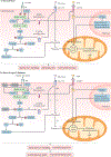
Fig. 5 |. Insulin signalling in the heart in normal conditions and in type 2 diabetes mellitus.
a | In normal conditions, insulin binds to the α-subunits of the insulin receptor (IR) in cardiomyocytes, which induces the phosphorylation of the IR β-subunits. This phosphorylation triggers the activation of the docking protein IR substrate 1 (IRS1), which subsequently activates phosphatidylinositol 3-kinase (PI3K) and RACβ serine/threonine-protein kinase 2 (AKT2), which has a critical role in glucose metabolism. PI3K and AKT2 activation promotes the translocation of glucose transporter 4 (GLUT4) and the free fatty acid (FFA) transporter CD36 from intracellular stores to the plasma membrane, thereby leading to increased glucose and FFA uptake. FFAs can activate the transcription factor peroxisome proliferator-activated receptor-α (PPARα), which induces the expression of multiple genes related to lipid metabolism. Increased glucose and FFA uptake increases mitochondrial oxidative metabolism to generate ATP via the tricarboxylic acid (TCA) cycle and β-oxidation, which supports myocardial contractile function. Insulin-mediated activation of AKT2 leads to inhibitory phosphorylation of glycogen synthase kinase 3β (GSK3β), which increases glycogen synthesis by glycogen synthase (GS). b| In type 2 diabetes mellitus, insulin signalling in cardiomyocytes is impaired. Signalling through the insulin-dependent glucose intake pathway is diminished, leading to increased FFA intake via CD36 and to eventual lipid accumulation. However, the excessive amount of FFA exceeds the capacity of mitochondrial respiration to generate ATP, leading to cardiomyocyte death, impaired cardiac function and lipid accumulation and toxicity. Potential therapeutic targets for metabolic disturbances that have been tested in preclinical models or in clinical studies are marked with an asterisk. HK, hexokinase; P, phosphorylation; PDK, pyruvate dehydrogenase kinase; PtdIns(4,5)P2, phosphatidylinositol (4,5)-bisphosphate; PtdIns(3,4,5)P3, phosphatidylinositol (3,4,5)-trisphosphate; PTEN, phosphatase and tensin homologue; PTP1B, protein tyrosine phosphatase 1B; TRB3, tribbles homologue 3.

References

    1. Rubler S. et al. New type of cardiomyopathy associated with diabetic glomerulosclerosis. Am. J. Cardiol. 30, 595–602 (1972). - PubMed
    1. Kannel WB, Hjortland M & Castelli WP Role of diabetes in congestive heart failure: the Framingham study. Am. J. Cardiol. 34, 29–34 (1974). - PubMed
    1. Tarquini R et al. Clinical approach to diabetic cardiomyopathy: a review of human studies. Curr. Med. Chem. 25, 1510–1524 (2018). - PubMed
    1. Bouthoorn S et al. The prevalence of left ventricular diastolic dysfunction and heart failure with preserved ejection fraction in men and women with type 2 diabetes: a systematic review and meta-analysis. Diab. Vasc. Dis. Res. 15, 477–493 (2018). - PMC - PubMed
    1. Dauriz M et al. Prognostic impact of diabetes on long-term survival outcomes in patients with heart failure: a meta-analysis. Diabetes Care 40, 1597–1605 (2017). - PubMed

Publication types

Substances