Review
. 2020 Apr;21(4):333-348.
doi: 10.1111/tra.12722. Epub 2020 Mar 1.

Fluorogen activating protein toolset for protein trafficking measurements

Affiliations
Review

Fluorogen activating protein toolset for protein trafficking measurements

Lydia A Perkins et al. Traffic. 2020 Apr.

Abstract

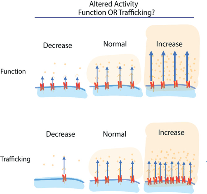
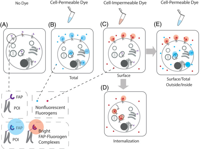
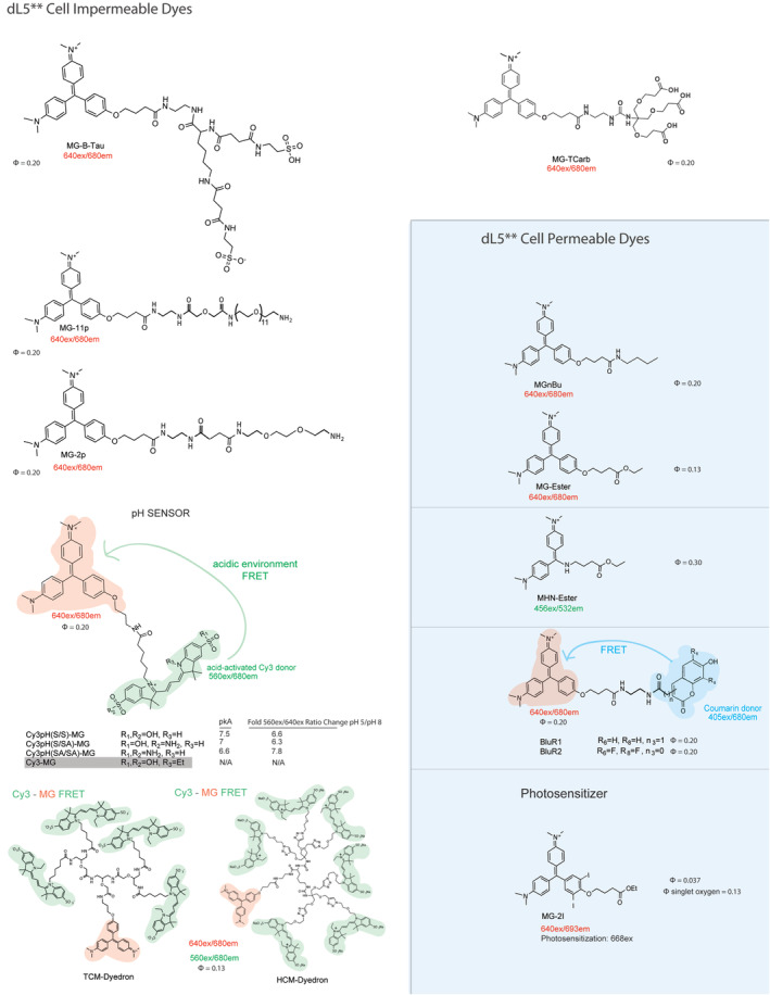
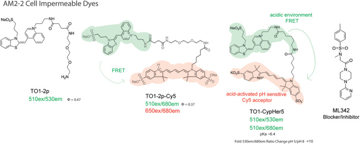
Throughout the past decade the use of fluorogen activating proteins (FAPs) has expanded with several unique reporter dyes that support a variety of methods to specifically quantify protein trafficking events. The platform's capabilities have been demonstrated in several systems and shared for widespread use. This review will highlight the current FAP labeling techniques for protein traffic measurements and focus on the use of the different designed fluorogenic dyes for selective and specific labeling applications.

Keywords: fluorogen activating proteins (FAP); fluorogenic dye; protein trafficking.


Conflict of interest statement

M.P.B. is founder and Chief Technology Officer at Sharp Edge Laboratories, Inc., a licensee commercially utilizing the FAP fluorogen technology.

Figures

Figure 1
Figure 1
Overall activity can be a result of altered protein function, altered protein expression at a target site (eg, the plasma membrane), or a combination of the two effects
Figure 2
Figure 2
A Snapshot of FAP tagged protein of interest (FAP‐POI) labeled with cell‐excluded or permeable dye without dye wash‐out post labeling. A, No Dye. Absence of fluorescence; B, Use of a cell‐permeant dye labels FAP‐tagged protein throughout the cell, including those in endocytic compartments, as well as those in biosynthetic compartments. This labeling gives a total protein fluorescence measurement. Alternatively (C) use of cell‐excluded dye selectively labels FAP‐tagged proteins at the plasma membrane (surface measurement), which may then be trafficked into the cell by endocytosis, allowing for measurements of internalization (D). E, Sequential labeling of the cells with the cell‐excluded dye followed by a cell permeant dye of the same or different spectral properties allows measurements of the fraction of total protein at the cell surface, at a single cell or population level, depending on the measurement technique
Figure 3
Figure 3
Fluorogenic dyes bound by dL5**. Cell‐excluded and permeable to the mammalian PM. MG2p and MG‐Ester have been used successfully with Mars1Cy and Mars1 FAPs23
Figure 4
Figure 4
Fluorogenic dyes specific for AM2‐2. Cell‐excluded to mammalian PM. There is no cell‐permeant TO1 analog due to residual DNA binding of the fluorogen within the cell. FRET and pH sensitive variants were developed using Cy5 as an acceptor, with binding selectivity but weak fluorogenic activation upon binding, due to the intrinsic transfer from the donor fluorogen even in the unbound state. Binding to cell surface receptors typically provides adequate concentration and activation in solution to conduct unwashed experiments measuring endocytosis of AM2‐2 labeled receptors using these dyes. The nonfluorescent inhibitor ML342 was identified as a potent inhibitor (approximately 2 nM EC50) of TO1‐2p binding to the AM2‐2 FAP
Figure 5
Figure 5
Other fluorogenic dyes that bind to different FAP scFVs
Figure 6
Figure 6
The right dyes at the right time for quantitative assessment of protein trafficking changes without need of wash‐steps. A and B, Real‐time cell surface protein trafficking measurements. C‐E, Detail end‐point trafficking measurements. A, Cell‐impermeable dye is added to the cell followed by drug, allowing for measurement of active trafficking itineraries. Both dL5** and AM2‐2 FAPs and their respective dyes will generate this measurement. B, The self‐checking internalization assay for AM2‐2 FAP consists of first addition of a cell‐excluded fluorogen of one color of moderate affinity, induction of internalization via drug addition, and then competitive displacement of remaining surface FAP‐fluorogen complex with a second cell‐excluded, high‐affinity and different color dye, followed by quantitative assessment of the color ratio. The second dye only labels receptors that are remaining at the plasma membrane, and internalized proteins are protected from displacement. Because all dyes are cell‐excluded, this labeling approach is selective for surface trafficking and does not label proteins contained within biosynthetic compartments.32 C, Drug is added to cells followed by cell‐impermeable dye for a surface FAP‐POI measurement to quantify cell surface POI trafficking changes. dL5**, AM2‐2, Mars1Cy FAPs and their respective dyes will generate this measurement. D, Drug is added to cells followed by cell‐permeable dye for total FAP‐POI measurement. Both dL5** and Mars1Cy FAPs and their respective dyes will generate this measurement. E, Drug is added to cells followed by cell‐impermeable dye for a surface FAP‐POI measurement. Subsequently, a cell‐permeable dye (same color or different color) is added and then total FAP‐POI is measured for the surface/total FAP‐POI. This is a dL5** and Mars1Cy FAP specific assay format, because the TO1 and other fluorogens are not available in cell permeant, highly specific forms
Figure 7
Figure 7
Diagram to show the signal changes related to different steps of Cy3(S/SA)pH‐MG labeled FAP‐B2AR internalization. MG signal (640ex/680em) stays consistent over the trafficking itinerary while pH sensitive FRET signal (560ex/680em) intensity is dependent on the local pH environment

References

    1. Rodriguez EA, Campbell RE, Lin JY, et al. The growing and glowing toolbox of fluorescent and photoactive proteins. Trends Biochem Sci. 2017;42(2):111‐129. 10.1016/j.tibs.2016.09.010. - DOI - PMC - PubMed
    1. Li C, Tebo AG, Gautier A. Fluorogenic labeling strategies for biological imaging. Int J Mol Sci. 2017;18(7):e1473 10.3390/ijms18071473. - DOI - PMC - PubMed
    1. Thorn K. Genetically encoded fluorescent tags. Mol Biol Cell. 2017;28(7):848‐857. 10.1091/mbc.E16-07-0504. - DOI - PMC - PubMed
    1. Jullien L, Gautier A. Fluorogen‐based reporters for fluorescence imaging: a review. Method Appl Fluoresc. 2015;3(4):042007 10.1088/2050-6120/3/4/042007. - DOI - PubMed
    1. Yan Q, Bruchez MP. Advances in chemical labeling of proteins in living cells. Cell Tissue Res. 2015;360(1):179‐194. 10.1007/s00441-015-2145-4. - DOI - PMC - PubMed

Publication types

LinkOut - more resources