. 2020 May;97(5):966-979.
doi: 10.1016/j.kint.2019.11.013. Epub 2019 Nov 28.

IL-6-mediated hepatocyte production is the primary source of plasma and urine neutrophil gelatinase-associated lipocalin during acute kidney injury

Affiliations

IL-6-mediated hepatocyte production is the primary source of plasma and urine neutrophil gelatinase-associated lipocalin during acute kidney injury

Nataliya I Skrypnyk et al. Kidney Int. 2020 May.

Abstract

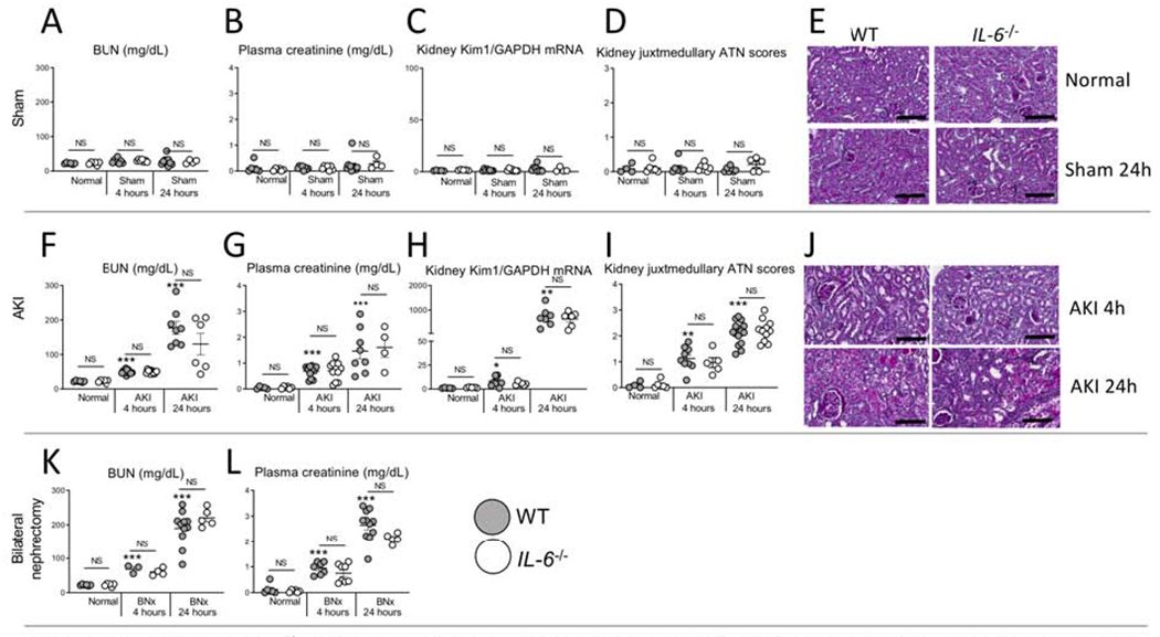
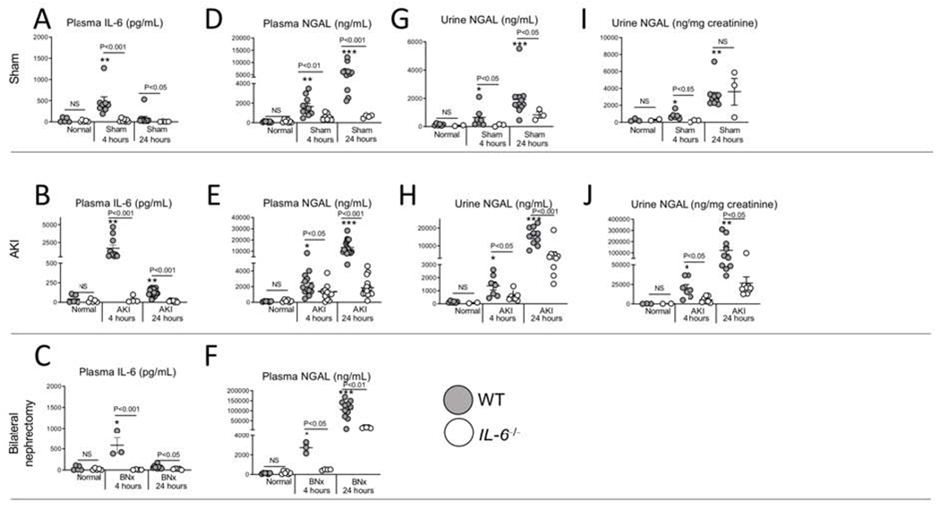
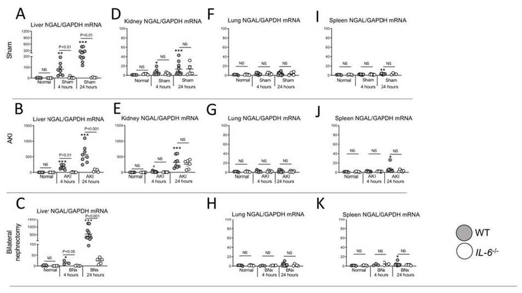
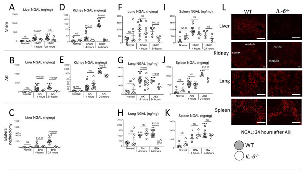
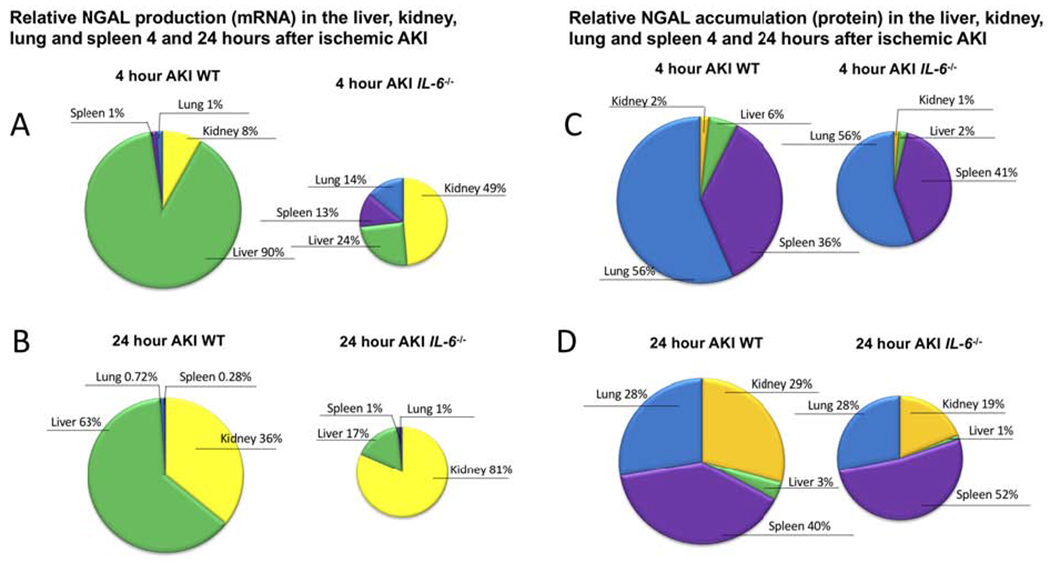
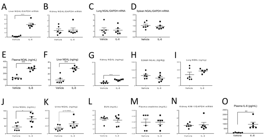
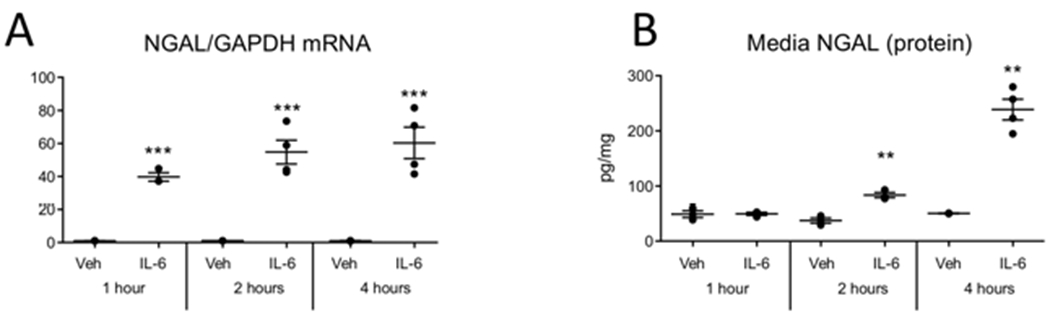
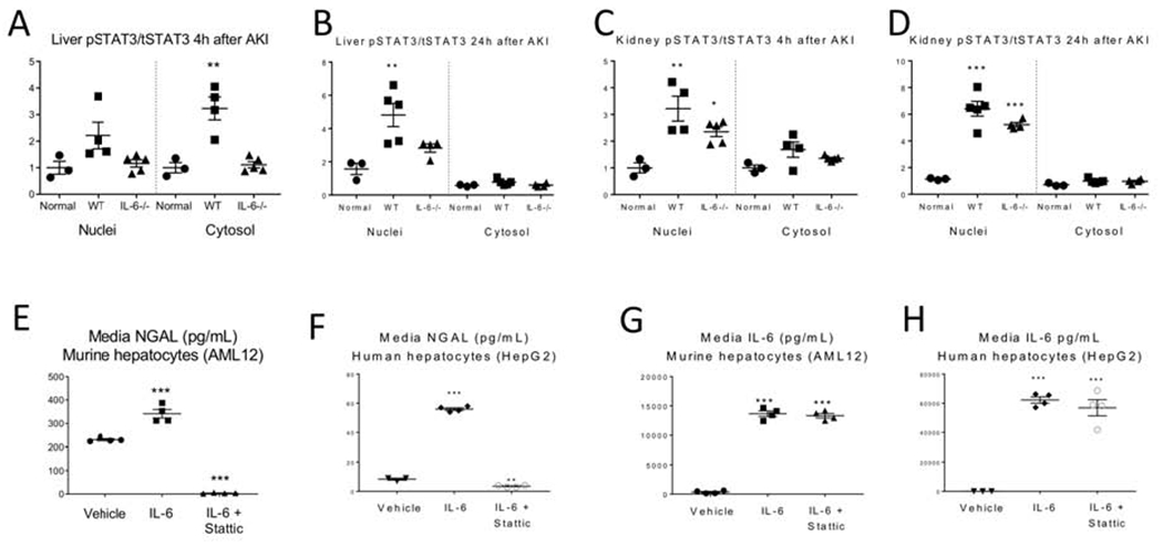
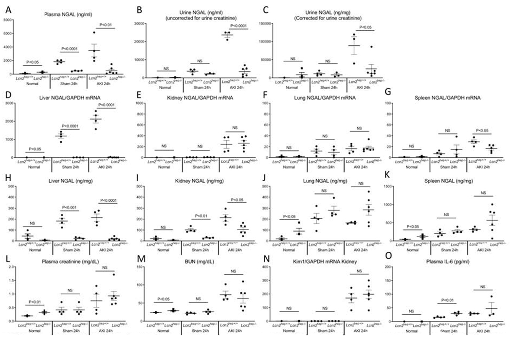
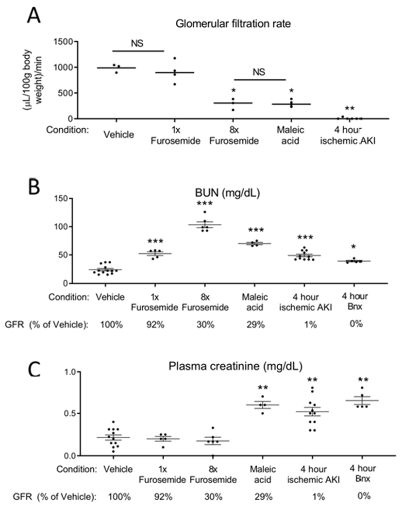
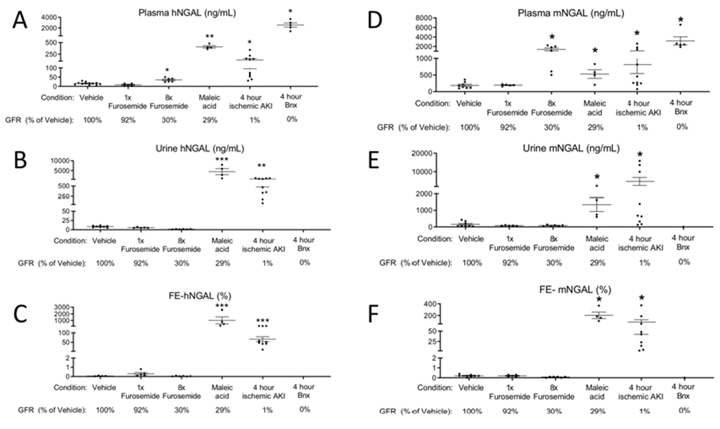
Neutrophil gelatinase associated lipocalin (NGAL, Lcn2) is the most widely studied biomarker of acute kidney injury (AKI). Previous studies have demonstrated that NGAL is produced by the kidney and released into the urine and plasma. Consequently, NGAL is currently considered a tubule specific injury marker of AKI. However, the utility of NGAL to predict AKI has been variable suggesting that other mechanisms of production are present. IL-6 is a proinflammatory cytokine increased in plasma by two hours of AKI and mediates distant organ effects. Herein, we investigated the role of IL-6 in renal and extra-renal NGAL production. Wild type mice with ischemic AKI had increased plasma IL-6, increased hepatic NGAL mRNA, increased plasma NGAL, and increased urine NGAL; all reduced in IL-6 knockout mice. Intravenous IL-6 in normal mice increased hepatic NGAL mRNA, plasma NGAL and urine NGAL. In mice with hepatocyte specific NGAL deletion (Lcn2hep-/-) and ischemic AKI, hepatic NGAL mRNA was absent, and plasma and urine NGAL were reduced. Since urine NGAL levels appear to be dependent on plasma levels, the renal handling of circulating NGAL was examined using recombinant human NGAL. After intravenous recombinant human NGAL administration to mice, human NGAL in mouse urine was detected by ELISA during proximal tubular dysfunction, but not in pre-renal azotemia. Thus, during AKI, IL-6 mediates hepatic NGAL production, hepatocytes are the primary source of plasma and urine NGAL, and plasma NGAL appears in the urine during proximal tubule dysfunction. Hence, our data change the paradigm by which NGAL should be interpreted as a biomarker of AKI.

Keywords: IL-6; acute kidney injury; biomarkers; cytokines; ischemia reperfusion; nephrotoxicity.


Figures

Figure 1.
Figure 1.. IL-6 deficient (IL-6−/−) mice have similar kidney function and injury after sham (surgery alone), ischemic acute kidney injury (AKI) or bilateral nephrectomy (BNx).
Kidney injury was determined in normal (unmanipulated) mice, and after sham, AKI or BNx at 4 and 24 hours in wild type (WT) and IL-6−/− mice. No kidney injury occurred after sham (surgery alone) in either WT or IL-6−/− as judged by (A) BUN, (B) plasma creatinine, (C) kidney KIM-1 mRNA, and (D) ATN score with (E) representative PAS images. Kidney injury was similar at 4 and 24 hours after AKI between WT and IL-6−/− mice as judged by (F) BUN, (G) plasma creatinine, (H) kidney mRNA KIM-1, and (I) ATN scores with (J) representative PAS images. Wild type and IL-6−/− mice with bilateral nephrectomy had similar levels of (K) BUN and (L) plasma creatinine. N=3 to 12 mice per group from 3 experiments. Results are expressed as mean ±SEM and analyzed by t test: WT versus IL-6−/− at the same time point (indicated above the bar) and versus normal WT (indicated below the bar: *P<0.05; **P<0.01, ***P < 0.0001). Scale bar: 100 μm.
Figure 2.
Figure 2.. Plasma NGAL is reduced in IL-6 deficient IL-6−/− mice after sham (surgery alone), ischemic acute kidney injury (AKI) or bilateral nephrectomy (BNx) at 4 and 24 hours post-procedure.
Plasma IL-6, plasma NGAL, and urine NGAL were determined in normal (unmanipulated) mice, and after sham, ischemic AKI or BNx at 4 and 24 hours in wild type (WT) and IL-6−/− mice. Plasma IL-6 was increased in WT 4 and 24 hours after (A) sham, (B) AKI, and (C) BNx. Plasma NGAL was significantly reduced in IL-6−/− at 4 and 24 hours after (D) sham, (E) AKI and (F) BNx. Urine NGAL was reduced after sham and ischemic AKI in IL-6−/− (G-J). N=3 to 12 mice per group from 3 experiments. Results are expressed as mean ±SEM and analyzed by t test: WT versus IL-6−/− at the same time point (indicated above the bar) and versus normal WT (indicated below the bar: *P<0.05; **P<0.01, ***P < 0.0001).
Figure 3.
Figure 3.. NGAL production (mRNA) occurs predominantly in the liver after sham (surgery alone}, ischemic acute kidney injury (AKI) and bilateral nephrectomy (BNx), and liver NGAL mRNA is reduced in IL-6−/− mice.
mRNA NGAL levels were determined in the (A-C) liver, (D, E) kidney, (F-H) lung, and (I-K) spleen in normal (unmanipulated) mice, and 4 and 24 hours after sham, AKI or BNx in wild type (WT) and IL-6−/− mice. N=3 to 12 mice per group from 3 experiments. Results are expressed as mean ±SEM and analyzed by t test: WT versus IL-6−/− at the same time point (indicated above the bar) and versus normal WT (indicated below the bar: *P<0.05; **P<0.01, ***P < 0.0001).
Figure 4.
Figure 4.. NGAL protein accumulates in the liver, lung, spleen, and kidney at 4 and 24 hours after sham (surgery alone), ischemic acute kidney injury (AKI) or bilateral nephrectomy (BNx).
NGAL protein levels (ELISA) were determined in normal (unmanipulated) mice, and 4 and 24 hours after sham, AKI and BNx in wild type (WT) and IL-6−/− mice in the (A-C) liver, (D-E) kidney, (F-H) lung, and (I-K) spleen. (L) Immunofluorescence staining for NGAL was performed 24 hours after AKI in WT and IL-6−/− in the liver, kidney, lung, and spleen (representative images). N=3 to 12 mice per group from 3 experiments. Results are expressed as mean ±SEM and analyzed by t test: WT versus IL-6−/− at the same time point (indicated above the bar) and versus normal WT (indicated below the bar: *P<0.05; **P<0.01, ***P < 0.0001). Scale bar: 100 μm.
Figure 5.
Figure 5.. Relative NGAL production (mRNA) and accumulation (protein) after ischemic AKI in wild type (WT) and IL-6−/− mice.
(A,B) NGAL production (mRNA) in liver, kidney, lung, spleen is expressed as % organ production and demonstrates that the liver is the major organ source of NGAL production 4 and 24 hours post-AKI in WT mice after ischemic AKI; in IL-6−/−, the kidney is the major source of NGAL production. Thus, IL-6 is a mediator of hepatic, but not renal, NGAL production. (C, D) NGAL protein levels in the liver, kidney, lung, and spleen are expressed as % organ levels and demonstrate that after AKI, NGAL accumulates predominantly within the kidney, lung, and spleen (total protein was determined within organs, thus, organ accumulation does not distinguish free NGAL from NGAL contained within neutrophils). A similar distribution of NGAL accumulation was observed in IL-6−/− mice. (The size of the pie chart were adjusted to indicate relative production and accumulation between WT and IL-6−/−.) The differences in mRNA and protein highlight that while the liver is the primary site of organ production, NGAL accumulation is widely distributed.
Figure 6.
Figure 6.. Intravenous (IV) administration of IL-6 to normal WT mice induces NGAL production (mRNA) in the liver and increases plasma NGAL.
200 ng of recombinant murine IL-6 or 0.1% BSA was administered IV every hour for 3 hours to healthy WT mice; endpoints were determined 1 hour after the last IV injection. mRNA NGAL expression was significantly increased in the (A) liver, but not the (B) kidney, (C) lung, or (D) spleen in IL-6-treated. Protein NGAL levels were significantly increased in the (E) plasma, (F) liver, and (G) kidney but not the (H) spleen, or (I) lung in IL-6-treated. Urine NGAL was significantly increased (J), but not when corrected for urine creatinine (K) in IL-6-treated. Kidney injury did not occur after IL-6 administration as judged by similar levels of (L) BUN, (M) plasma creatinine and (N) kidney KIM-1 mRNA expression. (O) Plasma levels of IL-6 were increased in mice receiving IV IL-6, as expected. Data were analyzed by t test, *P < 0.05, **P < 0.01 and ***P < 0.001, Vehicle vs IL-6. Results are expressed as mean ±SEM of expression values for 6 mice per group from 1 experiment, n=6).
Figure 7.
Figure 7.. IL-6 mediates NGAL production by 1 hour in hepatocytes, in vitro.
Vehicle (Veh) or 50 ng/mL of human recombinant IL-6 was added to human (HepG2) hepatocytes. (A) NGAL mRNA was increased in hepatocytes at 1, 2, and 4 hours after IL-6 addition. (B) NGAL protein levels in the media increased at 2 and 4, hours after IL-6 addition **P < 0.01 and ***P < 0.001 vs vehicle treated at the same time point by t test. n=2-5 for each condition.
Figure 8.
Figure 8.. IL-6 mediated NGAL production in the liver and hepatocytes is dependent on phosphorylation of STAT3.
Nuclear and cytosolic fractions were isolated from the liver and kidney from normal wild type (WT) mice and at 4 and 24 hours after AKI in WT and IL-6−/− mice and total and phosphorylated STAT3 (pSTAT3) was determined by ELISA. (A, B) In the liver. pSTAT3 increased and completely translocated to the nuclei at 24 hours in WT, but not IL-6−/− mice, with AKI. (C, D) In the kidney, pSTAT3 increased and translocated to the nucleus in both WT and IL-6−/− mice at 4 and 24 hours. IL-6 and an inhibitor of STAT3 phosphorylation (STATTIC) was added to murine (AML12) and human (HepG2) hepatocyte cell lines in vitro. IL-6 was added 1 hour after addition of Stattic; media NGAL was determined 4 hours after adding IL-6. (E-H) NGAL was increased with addition of IL-6 and reduced with IL-6+STATTIC in AML12 and HepG2. IL-6 was unchanged by STATTIC in AML12 and HepG2. Data represent the expression values for 3 to 5 mice per group from 1 experiment. Results are expressed as mean ±SEM. Data were analyzed by t test, *P < 0.05, **P < 0.01 and ***P < 0.001 vs. WT normal (uninjured) mice or vs. vehicle in cell culture experiments.
Figure 9.
Figure 9.. Hepatocyte-specific Lcn2 (NGAL) deficient mice have reduced plasma and urine NGAL after ischemic AKI.
Normal mice (no procedure), sham (surgery alone) and ischemic AKI were studied in hepatocyte specific Lcn2 deficient (Lcn2hep−/−) mice, Cre-negative floxed littermates were used as controls (Lcn2hep+/+). (A, B, C) Plasma and urine NGAL were dramatically reduced in Lcn2Hep−/− versus Lcn2Hep+/+. As expected, Liver mRNA NGAL expression was decreased in Lcn2hep−/− versus Lcn2Hep+/+; NGAL mRNA was similar in the (B) kidney and (C) lung in Lcn2Hep−/− versus Lcn2Hep+/+; (D) spleen mRNA NGAL was reduced after AKI in Lcn2Hep−/− versus Lcn2Hep+/+. NGAL protein levels were reduced in Lcn2hep−/− Lcn2Hep−/− versus Lcn2Hep+/+ in the (E) liver, and (K) kidney, but not the lung and spleen in AKI. Kidney function with AKI was similar as judged by (L) BUN, (M) plasma creatinine and (N) kidney mRNA KIM-1 expression. (O) Plasma 1L-6 levels were similar in AKI. Results are expressed as mean ±SEM; N = 4-6 mice per group from 3 exDeriments. Analyzed by t test. *P < 0.05. **P < 0.01 and ***P < 0.001, Lcn2hep+/+ vs. Lcn2hep−/−.
Figure 10.
Figure 10.. Ly6G+ cells are not the source of plasma or urine NGAL after ischemic acute kidney injury (AKI).
Normal mice (Nl), and mice with and without depletion of Ly6G+ cells were studied 4 and 24 hours after ischemic AKI. Ly6G is predominantly expressed on neutrophils. There was no change in kidney function/injury after AKI with Ly6G+ cell depletion as judged by (A) BUN, (B) plasma creatinine and (C) kidney Kim1 mRNA. Neither (D) plasma nor (E,F) urine NGAL were affected by Ly6G+ cell depletion. (G) Liver, (H) kidney, (I) spleen, and (J) lung NGAL production was not affected by Ly6G+ depletion. NGAL protein levels in the liver and kidney (K and L) were not affected by Ly6G+ cell deletion. NGAL protein was reduced in the spleen at 24 hours in the lung at 4 and 24 hours after AKI with Ly6G+ cell depletion (N). Neutrophil depletion was confirmed by flow cytometry of the spleen (O). Results are expressed as mean ±SEM; 2 separate experiments. Analyzed by t test, Ly6G+ versus Ly6G− at the same time point. (N=4-10)
Figure 11.
Figure 11.. Models of AKI.
(A) Transcutaneous GFR was measured in 5 groups: 1) mice treated with vehicle (saline), 2) mice treated with 0.5 mg furosemide (1x furosemide) to induce mild prerenal azotemia, 3) mice treated with 4 mg furosemide for 2 doses (8x furosemide) to induce severe prerenal azotemia, 4) mice treated with maleic acid to induce proximal tubular injury and Fanconi syndrome, and 5) mice subjected to ischemic AKI. GFR was determined 6 hours after vehicle, furosemide, and maleic acid treatment, and 4 hours after ischemic AKI. GFR was analyzed by one way ANOVA, comparing all groups. *P < 0.01 versus Vehicle and 1x furosemide, **P < 0.001 versus all other groups. Results are expressed as mean ±SEM. n=3-6. (B) BUN and (C) plasma creatinine were determined 6 hours after vehicle, 6 hours after 1x furosemide, 6 hours after 8x furosemide, 6 hours after maleic acid, 4 hours after ischemic AKI, and 4 hours after bilateral nephrectomy. GFR as a percentage of vehicle treated is indicated for reference (GFR was not measured in the bilateral nephrectomy group, but is assumed to be “0” since both kidneys are removed). BUN and plasma creatinine were analyzed by t test versus vehicle: *P<0.05; **P<0.001, ***P<0.0001. Results are expressed as mean ±SEM. n=5-12 (groups include mice that directly had GFR measured, as well as additional mice).
Figure 12.
Figure 12.. Intravenously administered recombinant human (h) NGAL increases in the urine of mice with maleic acid administration and ischemic AKI to a greater extant than during pre-renal azotemia (furosemide administration).
rhNGAL was administered intravenously(IV) 6h after vehicle, furosemide, and maleic acid; and 4 hours ischemic AKI and bilateral nephrectomy (BNx). Endpoints were determined 1 after IV hNGAL administration. (A) hNGAL was significantly increased in the plasma after severe prerenal azotemia (8x furosemide), maleic acid administration, ischemic AKI, and bilateral nephrectomy versus vehicle. (B) Urine hNGAL was significantly increased after maleic acid treatment and ischemic AKI versus vehicle. (C) Fractional excretion (FE) of hNGAL was less than 1% in vehicle, mild prerenal azotemia (1x furosemide), and severe prerenal azotemia (8x furosemide), and was greater than 10% after maleic acid treatment and ischemic AKI. (D-F) Endogenous murine (m) NGAL was determined in the plasma and urine, and FE-mNGAL was calculated. Analyzed by t test versus vehicle, *P<0.05, **P<0.01, ***P<0.001. n=5-12 (groups include mice that directly had GFR measured, as well as additional mice; GFR for each model relative to vehicle is indicated below for reference).

Comment in

  • AKI: an enlightening acronym with a shadow side.
    Zijlstra JG, van Meurs M, Moser J. Zijlstra JG, et al. Kidney Int. 2020 Jun;97(6):1301. doi: 10.1016/j.kint.2020.03.016. Kidney Int. 2020. PMID: 32444096 No abstract available.
  • The author replies.
    Faubel S. Faubel S. Kidney Int. 2020 Jun;97(6):1301-1302. doi: 10.1016/j.kint.2020.03.017. Kidney Int. 2020. PMID: 32444098 No abstract available.

References

    1. Palevsky PM, Zhang JH, O’Connor TZ, et al. Intensity of renal support in critically ill patients with acute kidney injury. The New England journal of medicine 2008; 359: 7–20. - PMC - PubMed
    1. American Society of Nephrology Renal Research Report. J Am Soc Nephrol 2005; 16: 1886–1903. - PubMed
    1. Nickolas TL, Schmidt-Ott KM, Canetta P, et al. Diagnostic and prognostic stratification in the emergency department using urinary biomarkers of nephron damage: a multicenter prospective cohort study. J Am Coll Cardiol 2012; 59: 246–255. - PMC - PubMed
    1. Parikh CR, Devarajan P, Zappitelli M, et al. Postoperative biomarkers predict acute kidney injury and poor outcomes after pediatric cardiac surgery. J Am Soc Nephrol 22: 1737–1747. - PMC - PubMed
    1. Parikh CR, Coca SG, Thiessen-Philbrook H, et al. Postoperative Biomarkers Predict Acute Kidney Injury and Poor Outcomes after Adult Cardiac Surgery. J Am Soc Nephrol 2011; 22: 1748–1757. - PMC - PubMed

Publication types