. 2020 Apr 17;432(9):3033-3049.
doi: 10.1016/j.jmb.2020.02.003. Epub 2020 Feb 17.

Conditional Disorder in Small Heat-shock Proteins

Affiliations

Conditional Disorder in Small Heat-shock Proteins

T Reid Alderson et al. J Mol Biol. .

Abstract

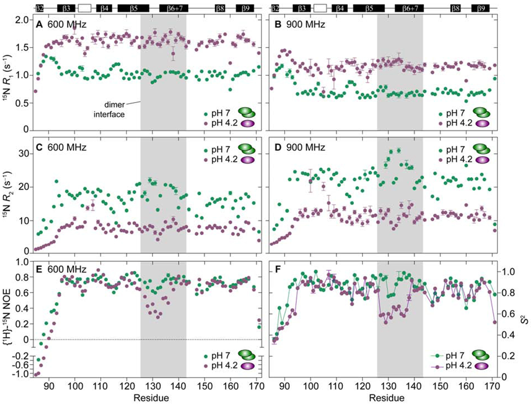
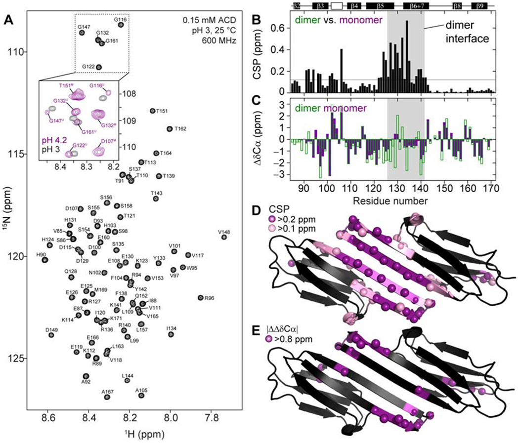
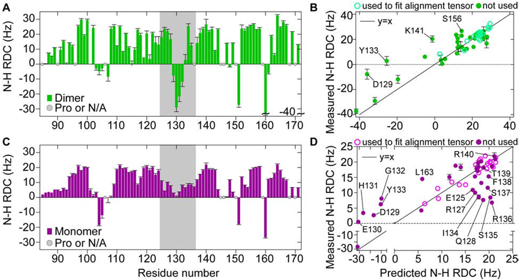
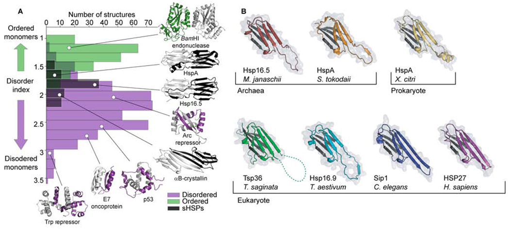
Small heat-shock proteins (sHSPs) are molecular chaperones that respond to cellular stresses to combat protein aggregation. HSP27 is a critical human sHSP that forms large, dynamic oligomers whose quaternary structures and chaperone activities depend on environmental factors. Upon exposure to cellular stresses, such as heat shock or acidosis, HSP27 oligomers can dissociate into dimers and monomers, which leads to significantly enhanced chaperone activity. The structured core of the protein, the α-crystallin domain (ACD), forms dimers and can prevent the aggregation of substrate proteins to a similar degree as the full-length protein. When the ACD dimer dissociates into monomers, it partially unfolds and exhibits enhanced activity. Here, we used solution-state NMR spectroscopy to characterize the structure and dynamics of the HSP27 ACD monomer. Web show that the monomer is stabilized at low pH and that its backbone chemical shifts, 15N relaxation rates, and 1H-15N residual dipolar couplings suggest structural changes and rapid motions in the region responsible for dimerization. By analyzing the solvent accessible and buried surface areas of sHSP structures in the context of a database of dimers that are known to dissociate into disordered monomers, we predict that ACD dimers from sHSPs across all kingdoms of life may partially unfold upon dissociation. We propose a general model in which conditional disorder-the partial unfolding of ACDs upon monomerization-is a common mechanism for sHSP activity.

Keywords: Conditional disorder; Molecular chaperone; NMR; Residual dipolar couplings; Small heat-shock protein.


Conflict of interest statement

Conflict of Interest

The authors declare no conflict of interest.

Figures

Fig. 1:
Fig. 1:. Domain architecture of HSP27 and NMR spectra of its ACD in various states.
(A) Linear domain depiction of HSP27 with numbers corresponding to domain boundaries. The predicted disorder based on the amino acid sequence is shown below, with values of 1 and 0 respectively corresponding to disordered and ordered. A horizontal line at 0.5 has been added for clarity. (B) Top: The cHSP27 construct contains the α-crystallin domain with the C137S mutation to prevent disulfide bond formation. Bottom: crystal structure of the dimeric ACD (PDB: 4mjh) with the β-strands β2 through β9 indicated. Right: cHSp27 exists as a dimer (green), monomers (purple), and an unfolded form (black). (C) The equilibrium arrows in panel B are strongly affected by pH. 2D 1H-N HSQC spectra of 2H, 13C, 15N-labeled cHSP27 as a function of pH. The percentage of dimer (D), monomer (M), and unfolded (U) are indicated in the lower-right of each spectrum. All spectra were recorded at 14.1 T and 25 °C and contoured at 10-fold the noise level (except panel D). Negative contours are shown in lighter colors in each spectrum. The insets depict a zoomed-in region. The single black contours in the pH 4.2 and 3.7 spectra highlight the resonances from the pH 3 sample (unfolded).
Fig. 2:
Fig. 2:. Structural changes upon ACD monomerization and unfolding.
(A) Projection of a 3D HNCA spectrum of 2H, 13C, 15N-labeled cHSP27 at pH 3 along the 1H and 15N dimensions. Resonance assignments are indicated. The inset contains overlaid spectra of cHSP27 at pH 3 (black) and pH 4.2 (purple), with resonance assignments for the unfolded state or monomer at pH 4.2 respectively indicated with a superscript U or M. Combined and weighted 13CO and 13Cα chemical shift perturbations (CSPs) for (B) the dimer at pH 7 vs. the monomer at pH 4.1. (C) Secondary 13Cα chemical shifts (Δδ13Cα) for the monomer (purple) at pH 4.1 and dimer (green) at pH 7. Negative values indicate β-strands. (D) CSPs from panel B mapped onto the structure of the cHSP27 dimer (PDB: 4mjh). The average value of the CSP is 0.11 ppm with the average plus one standard deviation equal to 0.23 ppm. (E) Residues with large changes (|Δδ13dimer - Δδ13monomer| > 0.8 ppm) are indicated on the structure of the cHSP27 dimer.
Fig. 3:
Fig. 3:. Backbone dynamics of the cHSP27 dimer and monomer.
15N spin relaxation data recorded on the cHSP27 dimer at pH 7 (green) and cHSP27 monomer at pH 4.2 (purple). 15N R1 rates recorded at a static magnetic field strength of (A) 600 and (B) 900 MHz, 15N R rates that were converted to R2 rates at (C) 600 and (D) 900 MHz, and {1H}15N NOEs measured at 600 MHz (E). Lipari-Szabo modelfree analysis of the 15N relaxation data from the dimer at pH 7 (green) and the monomer at pH 4.2 (purple). The squared generalized order parameters S2 are reported here (F).
Fig. 4:
Fig. 4:. Structural analysis of the cHSP27 dimer and monomer with RDCs.
1H-15N residual dipolar couplings (RDCs) for the cHSP27 dimer (A) or monomer (C) aligned in the presence of 4.3% (v/v) PEG- hexanol. The grey box indicates the region with large 13Cα and 13CO CSPs. RDCs from residues in well- defined regions of secondary structure were used to determine the alignment tensor that was then used to back-calculate predicted RDCs based on the crystal structure of the dimer (B) or one chain from the dimer (D). Outliers are indicated on the correlation plot. For the dimer, the outliers likely originate from structural noise in the 2.6-Å crystal structure. For the monomer, outliers largely cluster to the p5, L5,6+7, and β6+7 regions, indicative of a structural rearrangement relative to the crystal structure.
Fig. 5:
Fig. 5:. Partial unfolding upon ACD dimer dissociation is likely a general property.
(A) Disorder indices, calculated as the ratio of per-residue SASA to per-residue BSA of each protein in a complex, discriminate between dimers that dissociate into ordered (green) or disordered (purple) monomers. sHSP ACDs (black) largely overlap with the disordered monomers, suggesting that aCd monomers undergo at least partial unfolding upon dissociation. The structures depict examples from the various groups: BamHI endonuclease (1.12), HspA (3GLA; 1.67), Hsp16.5 (1SHS; 1.75), Arc repressor (1ARQ; 2.01), αB-crystallin (2KLR; 2.03), p53 TAD bound to NCBD (2L14; 2.52), HPV E7 CR3 domain (2B9D; 2.87), and the DNA-binding domain of Trp repressor (3WRP; 3.00). (B) Structures of ACD dimers from all kingdoms of life, only one chain from the dimer is shown here for clarity. The protein and organism names are listed under each structure.

References

    1. Haslbeck M, Weinkauf S, Buchner J, Small heat shock proteins: Simplicity meets complexity., J. Biol. Chem. 294 (2019) 2121–2132. doi:10.1074/jbc.REV118.002809. - DOI - PMC - PubMed
    1. Kampinga HH, de Boer R, Beerstra N, The multicolored world of the human HSPB family, Springer International Publishing, Cham, 2015. doi:10.1007/978-3-319-16077-1. - DOI
    1. Mogk A, Ruger-Herreros C, Bukau B, Cellular Functions and Mechanisms of Action of Small Heat Shock Proteins, Annu. Rev. Microbiol. 73 (2019) annurev-micro-020518–115515. doi :10.1146/annurev-micro-020518-115515. - DOI - PubMed
    1. Janowska MK, Baughman HER, Woods CN, Klevit RE, Mechanisms of Small Heat Shock Proteins, Cold Spring Harb. Perspect. Biol. (2019) a034025. doi:10.1101/cshperspect.a034025. - DOI - PMC - PubMed
    1. Arrigo AP, HSP27: novel regulator of intracellular redox state., IUBMB Life. 52 (2001) 303–07. doi:10.1080/152165401317291165. - DOI - PubMed

Publication types