. 2020 May 15:219:112847.
doi: 10.1016/j.physbeh.2020.112847. Epub 2020 Feb 18.

Sex-dependent effects of chronic variable stress on discrete corticotropin-releasing factor receptor 1 cell populations

Affiliations

Sex-dependent effects of chronic variable stress on discrete corticotropin-releasing factor receptor 1 cell populations

Zachary J Rosinger et al. Physiol Behav. .

Abstract

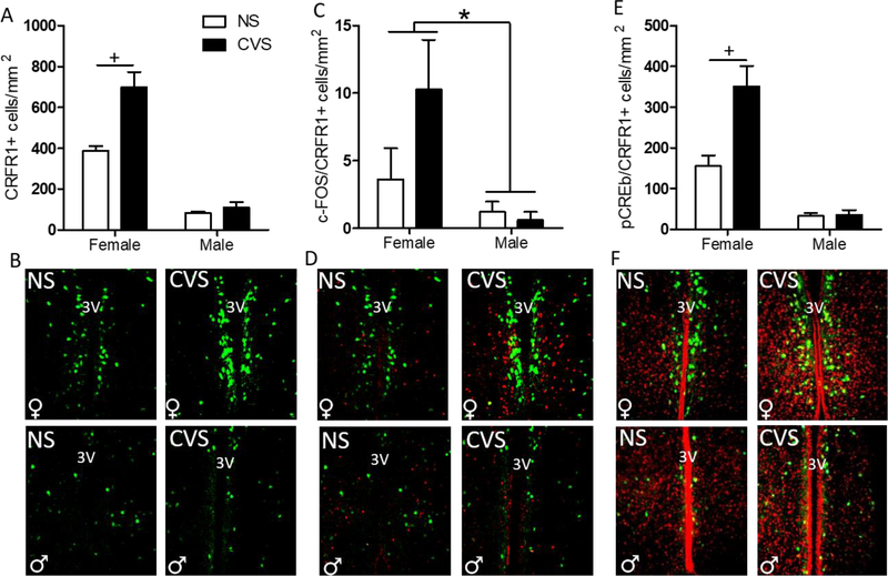
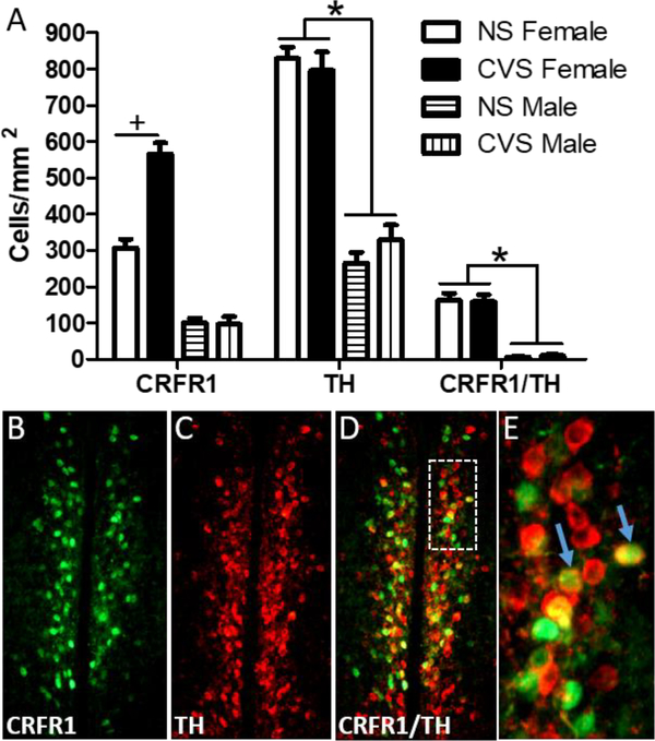
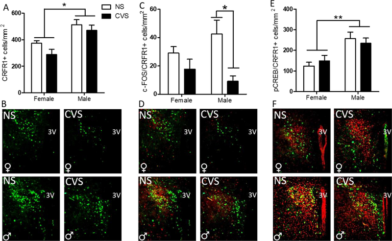
Anxiety and depression are strikingly more prevalent in women compared with men. Dysregulation of corticotropin-releasing factor (CRF) binding to its cognate receptor (CRFR1) is thought to play a critical role in the etiology of these disorders. In the present study, we investigated whether there were sex differences in the effects of chronic variable stress (CVS) on CRFR1 cells using CRFR1-GFP reporter mice experiencing a 9-day CVS paradigm. Brains were collected from CVS and stress naïve female and male mice following exposure to the open field test. This CVS paradigm effectively increased anxiety-like behavior in female and male mice. In addition, we assessed changes in activation of CRFR1 cells (co-localization with c-Fos and phosphorylated CREB (pCREB)) in stress associated brain structures, including two sexually dimorphic CRFR1 cell groups in the anteroventral periventricular nucleus (AVPV/PeN; F>M) and paraventricular hypothalamus (PVN; M>F). CVS increased CRFR1-GFP cell number as well as the number of CRFR1/pCREB co-expressing cells in the female but not male AVPV/PeN. In the PVN, the number of CRFR1/pCREB co-expressing cells was overall greater in males regardless of treatment and CVS resulted in a male-specific reduction of CRFR1/c-Fos cells. In addition, CVS induced a female-specific reduction in CRFR1/c-Fos cells within the anteroventral bed nucleus of the stria terminalis and both sexes exhibited a reduction in CRFR1/c-Fos co-expressing cells following CVS within the ventral basolateral amygdala. Overall, these sex-specific effects of CVS on CRFR1 populations may have implications for sex differences in stress-induction of mood disorders.

Keywords: Anxiety; Chronic stress; Corticotropin releasing factor; Sex difference.


Figures

Figure 1.
Figure 1.. Chronic variable stress (CVS) timeline.
The CVS timeline illustrates conditions to which CVS-exposed mice were subjected, and the final open field exposure that all mice, CVS and NS, were exposed to on day 10. On day 10, animals were euthanized at 90 minutes following the onset of the open field exposure.
Figure 2.
Figure 2.. Anatomical locations within which CRFR1-GFP and co-labeled cells were quantified.
Plate numbers correspond to locations determined by the Allen Mouse Brain Reference Atlas (https://mouse.brain-map.org/static/atlas). Approximate ROI shapes for the AVPV/PeN (A), BSTdl (B, top), BSTav (B, bottom), PVN (C), CeA (D, top), BLAv (D, bottom), and ARC (E). AC; anterior commissure, OT; optic tract.
Figure 3.
Figure 3.. Effects of CVS on anxiety-like behavior in the open field.
On day 10 following CVS onset, male and female CVS and non-stressed mice were exposed to a final open field exposure within which anxiety-like behavior was assessed. (A) The latency to first entry, was significantly longer for CVS animals than non-stressed animals. (B) Total number of center entries, showing that CVS mice made significantly fewer center entries than non-stressed counterparts. (C) The total amount of time spent in the center was significantly reduced in CVS-exposed compared to non-stressed mice. (D) No significant group differences were found for total distance traveled. * Main effect of treatment, p < 0.05.
Figure 4.
Figure 4.. Effects of CVS on the number of AVPV/PeN CRFR1-GFP, c-Fos/CRFR1, and pCREB/CRFR1 cells.
(A) CVS females showed a significant increase in CRFR1-ir compared to all other treatment groups. (B) Representative images of CVS and NS female and male AVPV/PeN CRFR1-GFP. (C) c-Fos/CRFR1-GFP co-expression was significantly higher in all female AVPV/PeN than male, regardless of treatment condition. (D) Representative images of CVS and NS female and male c-Fos/CRFR1-GFP showing a female-specific reduction in c-Fos/CRFR1-GFP cells. (E) Females, regardless of treatment, had increased pCREB/CRFR1-GFP in the AVPV/PeN compared to males, and CVS females specifically had higher pCREB/CRFR1-GFP than any other groups. (F) Representative images of NS and CVS female and male pCREB/CRFR1-GFP in the AVPV/PeN showing female specific increased pCREB. 3V, third ventricle; NS, non-stressed; CVS, chronic variable stress. * Indicates a main effect of sex, (F>M; p < 0.05). + indicates a significant increase in CVS compared to NS females (p < 0.01).
Figure 5.
Figure 5.. Co-localization of AVPV CRFR1-GFP with TH following CVS.
(A) The number of CRFR1/TH co-localized cells was unaffected in male and female mice following CVS. This indicates that the increase in CRFR1-GFP labeled cells in CVS females occurs in an independent set of neurons. (B-D) Representative images of CRFR1-GFP, TH, and a merged image from a CVS female mouse, respectively. White inset box (D) indicates the area further magnified in (E). Arrows show examples of co-labeled neurons. * indicates a greater number of CRFR1-GFP, TH, and co-labeled cells in females compared to males, p < .001. + indicates p < .001 (NS compared to CVS females).
Figure 6.
Figure 6.. Effects of CVS on the number of PVN CRFR1-GFP, c-Fos/CRFR1, and pCREB/CRFR1 cells.
(A) The female PVN had significantly less CRFR1-GFP than the male, regardless of treatment, with no effect of CVS on CRFR1-GFP in either sex. (B) Representative PVN CRFR1-GFP images for NS and CVS female and male mice. (C) CVS males had significantly fewer c-Fos/CRFR1 co-localized cells than non-stressed male counterparts, with no differences between female groups. (D) Representative c-Fos/CRFR1-GFP labeling in female and male mice from NS and CVS treatments. (E) Males had an overall greater amount of pCREB/CRFR1 co-labeled cells than females, regardless of treatment condition, with representative PVN pCREB/CRFR1 images from NS and CVS female and male mice shown in (F). NS, non-stressed; CVS, chronic variable stress. Data are presented as mean ± SEM, and significance threshold set to p<0.05. * Indicates statistical significance p<0.05. ** indicates p<.01.
Figure 7.
Figure 7.. Effects of CVS on CRFR1-GFP cell number and co-localization with c-Fos and pCREB in various brain regions.
CRFR1-GFP, CRFR1/c-Fos, and CRFR1/pCREB co-labeled cells were quantified within the anteroventral portion of the bed nucleus of the stria terminalis (BSTav; A-C), dorsolateral portion of the bed nucleus of the stria terminalis (BSTdl; D-F), central amygdala (CeA; G-I), ventral basolateral amygdala (BLAv; J-L), and arcuate nucleus (ARC; M-O). CVS selectively reduced c-Fos/CRFR1-GFP co-labeled cells in female but not male BSTav (B). In the BLAv, the number of CRFR1/c-Fos co-labeled cells was significantly reduced in both sexes (K). There was also a trend toward a reduction in c-Fos/CRFR1 co-labeled cells in CVS mice within the arcuate nucleus (p=0.056; N), with CVS animals regardless of sex, trending toward reduced co-expression. No significant differences were found within the BSTdl or CeA. Data are reported as mean cells/mm2 ± SEM. + Indicates main effect of stress on outcome, p≤0.05. * Indicates an effect of sex on outcome, p≤0.05. & indicates a trend toward main effect of treatment, p = 0.056. NS, non-stressed; CVS, chronic variable stress.
Figure 8.
Figure 8.. Correlations between time spent in the center area of the open field and CRFR1-GFP, CRFR1/c-Fos, and CRFR1/pCREB labeled cells within various brain regions in female (top) and male (bottom) mice.
Dark gray cells indicate a significant correlation (p<.05). A positive number indicates a positive association between time spent in the center area and the number of CRFR1-GFP, CRFR1/c-Fos, and CRFR1/pCREB labeled cells. This analysis reveals correlations with behavior that occur in both sexes (CRFR1/c-Fos in the BSTav and BLAv), only in females (CRFR1 and CRFR1/pCREB in AVPV/PeN, CRFR1 in CeA), and only in males (CRFR1/c-Fos in PVN). R1; CRFR1.

References

    1. Aguilera G (1994), Regulation of pituitary ACTH secretion during chronic stress. Front Neuroendo 15:321–350. - PubMed
    1. Ahn S, Olive M, Aggarwal S, Krylove D, Ginty DD, Vinson C (1998), A dominant-negative inhibitor of CREB reveals that it is a general mediator of stimulus-dependent transcription of c-fos. Mol Cell Bio 18(2):967–977. - PMC - PubMed
    1. Algamal M, Ojo JO, Lungmus CP, Muza P, Cammarata C, Owens MJ, Mouzon BC, Diamond DM, Mullan M, Crawford F (2018), Chronic Hippocampal Abnormalities and Blunted HPA Axis in an Animal Model of Repeated Unpredictable Stress. Front Behav Neurosci 12:150. - PMC - PubMed
    1. Anisman H, Prakash P, Merali Z, Poulter MO (2007), Corticotropin releasing hormone receptor alterations elicited by acute and chronic unpredictable stressor challenges in stressor-susceptible and resilient strains of mice. Behav Brain Res 181(2):180–90. - PubMed
    1. Bailey KR, Crawley JN. Anxiety-Related Behaviors in Mice (2009). In: Buccafusco JJ, editor. Methods of Behavior Analysis in Neuroscience. 2nd edition. Boca Raton (FL): CRC Press/Taylor & Francis; Chapter 5.

Publication types

Substances