. 2020 Feb 21;11(2):142.
doi: 10.1038/s41419-020-2333-3.

UHRF1-repressed 5'-hydroxymethylcytosine is essential for the male meiotic prophase I

Affiliations

UHRF1-repressed 5'-hydroxymethylcytosine is essential for the male meiotic prophase I

Hongjie Pan et al. Cell Death Dis. .

Abstract

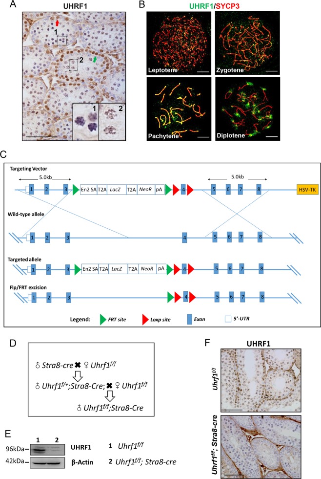
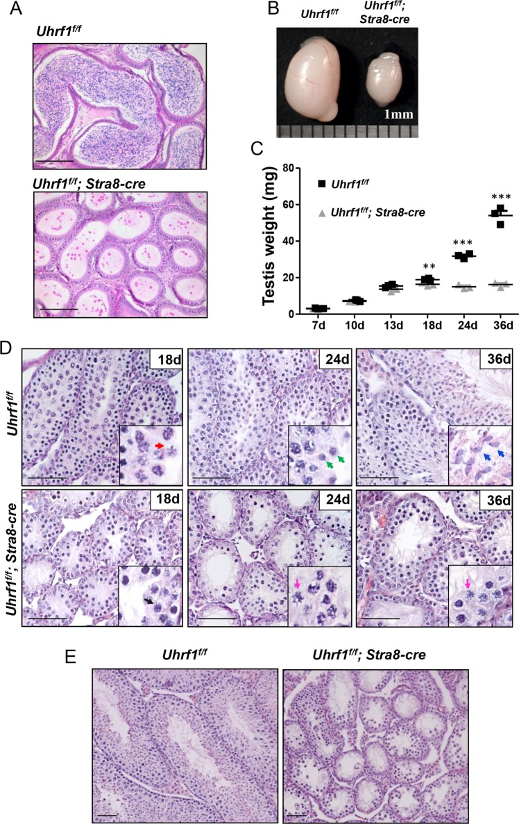
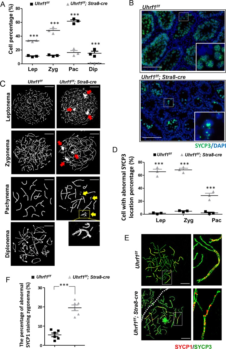
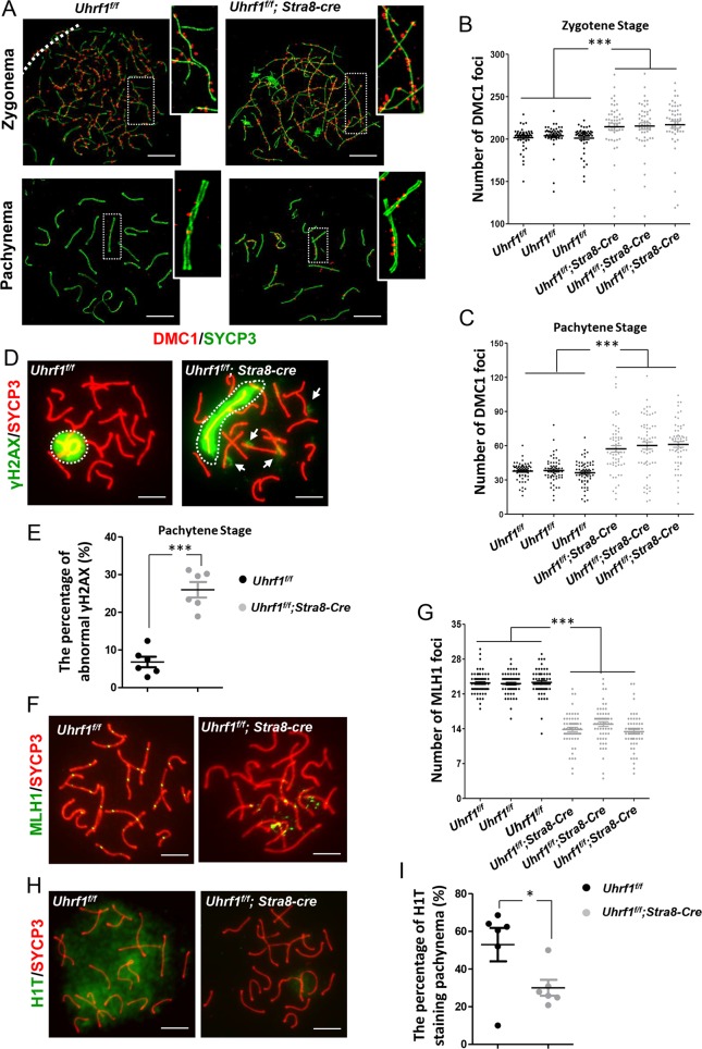
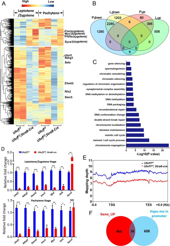
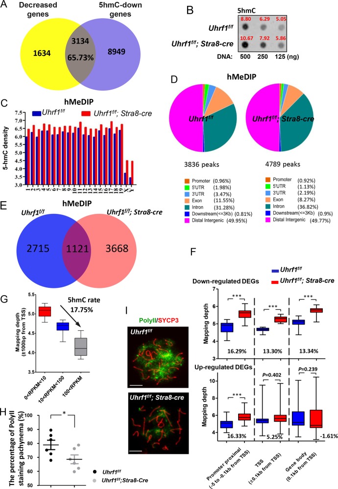
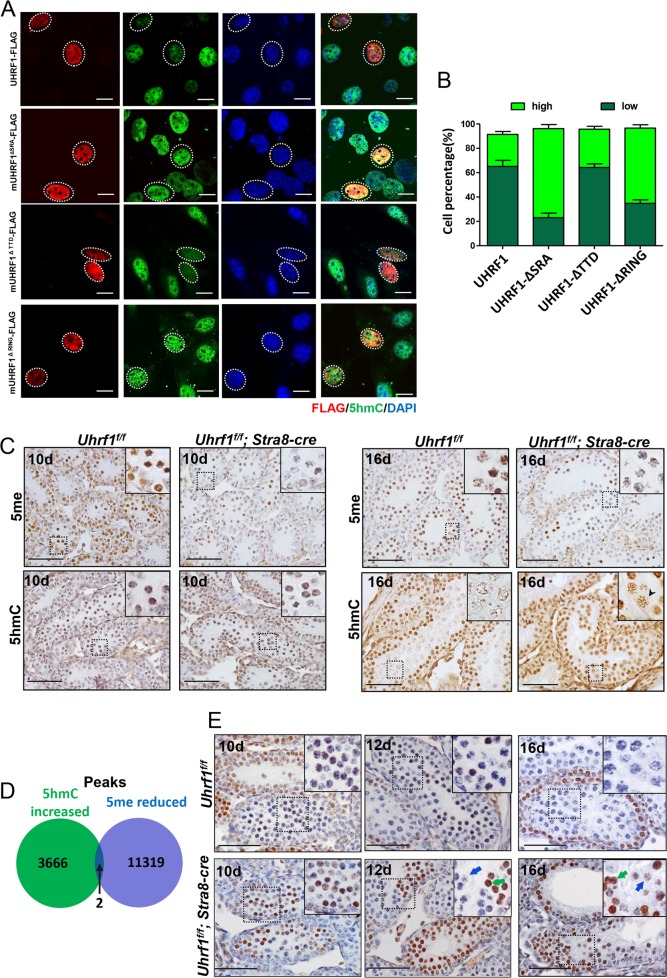
5'-hydroxymethylcytosine (5hmC), an important 5'-cytosine modification, is altered highly in order in male meiotic prophase. However, the regulatory mechanism of this dynamic change and the function of 5hmC in meiosis remain largely unknown. Using a knockout mouse model, we showed that UHRF1 regulated male meiosis. UHRF1 deficiency led to failure of meiosis and male infertility. Mechanistically, the deficiency of UHRF1 altered significantly the meiotic gene profile of spermatocytes. Uhrf1 knockout induced an increase of the global 5hmC level. The enrichment of hyper-5hmC at transcriptional start sites (TSSs) was highly associated with gene downregulation. In addition, the elevated level of the TET1 enzyme might have contributed to the higher 5hmC level in the Uhrf1 knockout spermatocytes. Finally, we reported Uhrf1, a key gene in male meiosis, repressed hyper-5hmC by downregulating TET1. Furthermore, UHRF1 facilitated RNA polymerase II (RNA-pol2) loading to promote gene transcription. Thus our study demonstrated a potential regulatory mechanism of 5hmC dynamic change and its involvement in epigenetic regulation in male meiosis.


Conflict of interest statement

The authors declare that they have no conflict of interest.

Figures

Fig. 1
Fig. 1. UHRF1 was expressed in the meiotic prophase I and the gene-trapped Uhrf1 conditional knockout mouse was prepared.
a The expression of UHRF1 protein in the mouse testicular tissue. b The distribution of UHRF1 protein (green) in the sub-stages of mouse meiotic prophase. c Schematic diagram of lox P-deletion system in the Uhrf1 gene in mouse; d mouse strategy; e, f western blot and immunohistochemical assay to the 16 dpp mouse testicle tissue showing the knockout efficiency of UHRF1 protein. At least three independent experiments were carried out. Scale bar, 25 μm in a, f, 5 μm in b.
Fig. 2
Fig. 2. UHRF1 was required for the mouse spermatogenesis and fertility.
a H&E staining of epididymal sections from 12-week-old Uhrf1f/f;Stra8-cre and Uhrf1f/f mice. b Morphology of testes. c The mean testicular weights of the mice from 7 to 36 dpp. d H&E staining of testicular sections at 18, 24, and 36 dpp. e Histology assay showing the testicular tubes in the adult Uhrf1f/f;Stra8-cre and Uhrf1f/f sections. Scale bar, 25 μm. dpp day post-partum. At least three independent experiments were carried out, data are presented as mean ± SEM of three mice in c. ***p ≤ 0.001; **p ≤ 0.01; NS, p > 0.05.
Fig. 3
Fig. 3. UHRF1 deletion disrupted the meiotic progression and synaptonemal complex assembly.
a Relative amounts of four spermatocyte populations (leptotene stage, zygotene stage, pachytene stage, and diplotene stage) during the prophase I in testes based on analyzing >600 spermatocytes in each stage. b, c The immunostaining of SYCP3 in the testicular sections (b) and surface-spread chromatin preparations of Uhrf1 deletion and control mice (c); d the percentage of spermatocytes with abnormal SYCP3 location. e Double immunofluorescence of testicular spread preparations of the adult mice, SYCP3 (green) and SYCP1 (red). f The percentage of spermatocytes with abnormal SYCP1 location. Lep leptotene, Zyg zygotene, Pac pachytene, Dip diplotene. Data are presented as mean ± SEM of three mice. ***p ≤ 0.001. Scale bar, 25 μm in b, 5 μm in c, e.
Fig. 4
Fig. 4. UHRF1 deficiency resulted in impaired meiotic recombination and defective pachynema.
a Double immunofluorescence of SYCP3 (green) and DMC1 (red) in testicular spread preparations. b, c The number of DMC1 foci in zygotene stage (b) and pachytene stage (c). d Immunostaining for SYCP3 (red) and γH2AX (green). e The percentage of abnormal γH2AX foci in the pachytene stage. f Immunostaining for SYCP3 (red) and MLH1 (green). g The number of MLH1 foci in pachynema. h Immunostaining for SYCP3 (red) and H1t (green). i The percentage of spermatocytes with H1T staining. ***p ≤ 0.001; *p ≤ 0.05. Scale bar, 5 μm in a, d, f, h.
Fig. 5
Fig. 5. The gene expression and global methylation after UHRF1 deletion.
a RNA-seq for mouse spermatocytes (leptotene/zygotene and pachytene stages). Clustered heat map of log2-transformed RPKMs showing the differentially expressed genes after UHRF1 deletion. Indicated genes are marked in right. b Identified differentially expressed genes were highly consistent between leptotene/zygotene and pachytene stages. (P pachytene stage genes, L leptotene/zygotene stage genes). c Gene ontology analysis of DEGs using the database of ClusterProfiler V3.4.4. d RT-PCR assay showing the relative change of some representative meiotic genes. e DNAme densities across the gene bodies of all reference genes. f Venn diagram depicting a set of 16 genes that were induced in transcripts and hypomethylated in promoter and 5’ UTR regions (unique in control) from the leptotene to pachytene stage in the UHRF1-deficient spermatocytes. For the upregulated transcripts, the cut-off was p value < 0.001 with fold change >2.
Fig. 6
Fig. 6. Hyper-5hmC resulted from UHRF1 deletion.
a Venn diagram depicting reduced transcripts associated with 5hmC downregulation. b 5hmC level of meiosis prophase I spermatocytes (16 dpp). c 5hmC densities of all chromosomes. d The distribution of 5hmC density on the genome of spermatocytes. e Venn diagram depicting 5hmC peaks in Uhrf1f/f;Stra8-cre and Uhrf1f/f spermatocytes. f 5hmC densities was shown in the proximal promoter, TSS, and gene body regions of the DEGs. g 5hmC densities in TSSs of the total refgenes with different RPKMs. h The percentage of RNA polymerase II staining in pachynema. i Double immunofluorescence of testicular spread preparations, SYCP3 (red) and RNA polymerase II (green). Scale bar, 5 μm in i.
Fig. 7
Fig. 7. The levels of 5hmC and TET1 were upregulated after hypo-DNAme.
a Overexpression of UHRF1, mUHRF1-ΔSRA-FLAG, mUHRF1-ΔTTD-FLAG, and mUHRF1-ΔRING-FLAG plasmids and the level of 5hmC in GC1 cells. b Percentage of the cells with strong or weak intensity of green fluorescence. c Immunohistochemistry assay of DNAme and 5hmC in the mouse spermatocytes. d Venn diagram showing the overlap between hyper-5hmC and hypo-DNAme peaks. The significance was evaluated by Fisher’s exact test. e Immunohistochemistry assay of TET1 in the mouse testis. Scale bar, 10 μm in a, 25 μm in c, e.

Similar articles

Cited by

References

    1. Guichaoua MR, et al. Meiotic anomalies in infertile men with severe spermatogenic defects. Hum. Reprod. 2005;20:1897–1902. doi: 10.1093/humrep/deh868. - DOI - PubMed
    1. Hann MC, Lau PE, Tempest HG. Meiotic recombination and male infertility: from basic science to clinical reality? Asian J. Androl. 2011;13:212–218. doi: 10.1038/aja.2011.1. - DOI - PMC - PubMed
    1. Kota SK, Feil R. Epigenetic transitions in germ cell development and meiosis. Dev. Cell. 2010;19:675–686. doi: 10.1016/j.devcel.2010.10.009. - DOI - PubMed
    1. Kaneda M, et al. Essential role for de novo DNA methyltransferase Dnmt3a in paternal and maternal imprinting. Nature. 2004;429:900–903. doi: 10.1038/nature02633. - DOI - PubMed
    1. Zamudio N, et al. DNA methylation restrains transposons from adopting a chromatin signature permissive for meiotic recombination. Genes Dev. 2015;29:1256–1270. doi: 10.1101/gad.257840.114. - DOI - PMC - PubMed

Publication types

MeSH terms