. 2020 Feb 21;10(1):3173.
doi: 10.1038/s41598-020-60120-4.

Systematic analysis of the Hippo pathway organization and oncogenic alteration in evolution

Affiliations

Systematic analysis of the Hippo pathway organization and oncogenic alteration in evolution

Yuxuan Chen et al. Sci Rep. .

Abstract

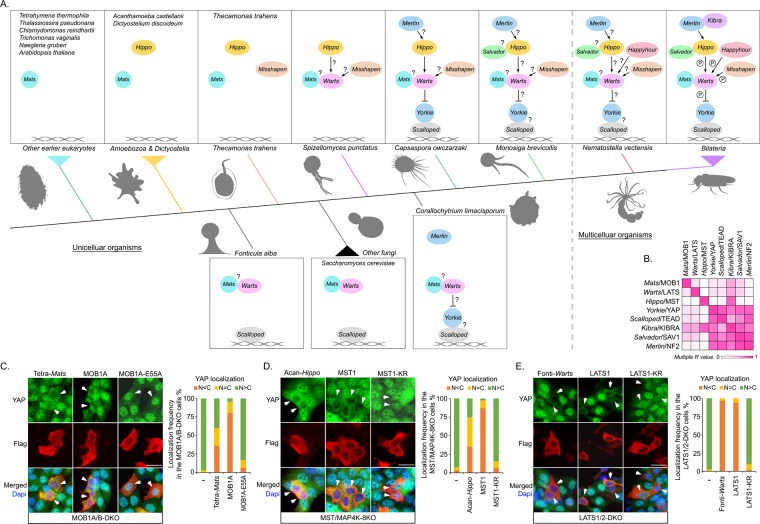
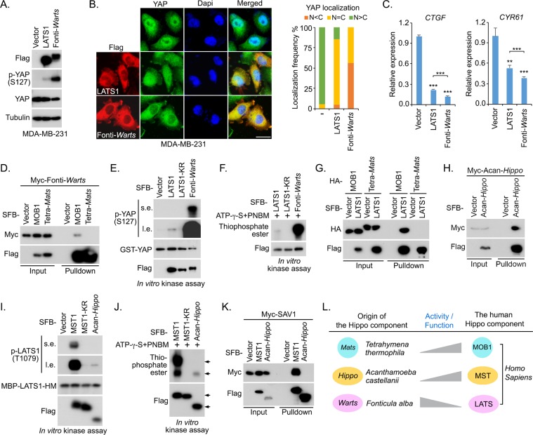
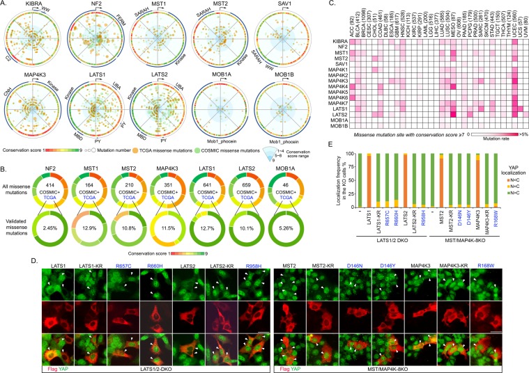
The Hippo pathway is a central regulator of organ size and a key tumor suppressor via coordinating cell proliferation and death. Initially discovered in Drosophila, the Hippo pathway has been implicated as an evolutionarily conserved pathway in mammals; however, how this pathway was evolved to be functional from its origin is still largely unknown. In this study, we traced the Hippo pathway in premetazoan species, characterized the intrinsic functions of its ancestor components, and unveiled the evolutionary history of this key signaling pathway from its unicellular origin. In addition, we elucidated the paralogous gene history for the mammalian Hippo pathway components and characterized their cancer-derived somatic mutations from an evolutionary perspective. Taken together, our findings not only traced the conserved function of the Hippo pathway to its unicellular ancestor components, but also provided novel evolutionary insights into the Hippo pathway organization and oncogenic alteration.


Conflict of interest statement

The authors declare no competing interests.

Figures

Figure 1
Figure 1
The development of the Hippo pathway paralogous genes is caused by the whole genome duplication in fish. (This figure is related to Fig. S1 and Tables S1–S3). (A) Illustration of the Hippo pathway paralogous genes in the indicated species from Drosophila to human. The Hippo pathway components were searched in the indicated species’ genomes by TBLASTN. The Hippo paralogous gene number in each indicated specie is shown. The listed species in Protostomia, Echinodermata and Leptocardii were labeled in grey; the listed species in Fish were labeled in pink; the listed species in Terapod were labeled in light blue. (B) The development of the Hippo pathway functional paralogous genes was a result of whole genome duplication. The q scores among the Hippo pathway functional paralogous genes were more significant than those of control genes. (C) The Hippo pathway is enriched with the duplicate genes saved after whole genome duplication as compared with the whole human genome. (D) Genomic regions near YAP and TAZ genes in both Danio rerio and Homo sapiens show a strong syntenic relationship. The neighboring gene information of YAP and TAZ were obtained from BioMart.
Figure 2
Figure 2
The Hippo pathway unicellular ancestor components show conserved activities in human cells. (This Figure is related to Figs. S2 and S3; Tables S1 and S4). (A) Schematic illustration of the Hippo pathway evolution from unicellular organisms to Bilateria. The Hippo pathway components were searched from the genomes of the indicated species ranging from Tetrahymena thermophila to Bilateria by TBLASTN. The color of lines was matched with the firstly emerged Hippo pathway component in the indicated species. The presumed complex formation and regulation were indicated by a question mark. The silhouette figures were made by the authors or obtained from http://phylopic.org. (B) Coevolution analysis among the Hippo pathway core components. Linear regression analysis was performed to determine the correlation between pairwise evolutionary distances based on a multiple protein sequence alignment. (C–E) Three Hippo pathway ancestor components show conserved functions in the human cells. The Hippo pathway unicellular ancestor genes Mats (from Tetrahymena thermophile), Hippo (from Acanthamoeba castellanii) and Warts (from Fonticula alba) were synthesized and respectively expressed in the MOB1A/B double knockout (DKO) (C), MST/MAP4K-8KO (D) and LATS1/2 DKO (E) HEK293A cells. Immunofluorescence was performed using YAP and Flag antibodies. The human MOB1A, MST1 and LATS1 were taken as positive controls, while their inactive mutant MOB1A-E55A, MST1 kinase-dead mutant (K59R) and LATS1 kinase-dead mutant (K734R) were included as negative controls. Flag-positive cells (arrows) from ~30 different views (~200 cells in total) were randomly selected and quantified for YAP localization. Scale bar, 40 μm.
Figure 3
Figure 3
Characterization of the Hippo pathway unicellular ancestor components. (This figure is related to Figs. S2–S4). (A–C) Fonticula alba Warts (Fonti-Warts) has a stronger ability than human LATS1 to phosphorylate YAP at S127 (A), translocate YAP into the cytoplasm (B) and inhibit YAP downstream gene transcription (C) in MDA-MB-231 cells. Flag-positive cells (~200 cells in total) were randomly selected and quantified for YAP localization. **p < 0.01, ***p < 0.001 (Student’s t-test). Scale bar, 40 μm. (D) Fonti-Warts interacts with human MOB1. HEK293T cells were transfected with the indicated constructs and subjected to the pulldown assay. (E and F) Fonti-Warts has a stronger kinase activity than human LATS1. SFB-LATS1, SFB-LATS1 kinase-dead mutant (LATS1-KR) and SFB-Fonti-Warts were purified from HEK293T cells by using S protein beads and washed thoroughly with high-salt buffer containing 250 mM NaCl. In vitro kinase assay was performed to examine their abilities to phosphorylate the bacterially purified GST-YAP (E) or induce the auto-phosphorylation (F). s.e., short exposure. l.e., long exposure. (G) Tetrahymena thermophile Mats (Tetra-Mats) failed to bind human LATS1. HEK293T cells were transfected with the indicated constructs and subjected to the pulldown assay. (H) Acanthamoeba castellanii Hippo (Acan-Hippo) can form as a dimer. HEK293T cells were transfected with the indicated constructs and subjected to the pulldown assay. (I,J) Acan-Hippo has a weaker kinase activity than human MST1. SFB-MST1, SFB-MST1 kinase-dead mutant (MST1-KR) and SFB-Acan-Hippo was purified from HEK293T cells by using S protein beads and washed thoroughly with high-salt buffer containing 250 mM NaCl. In vitro kinase assay was performed to examine their abilities to phosphorylate the bacterially purified MBP-LATS1 hydrophobic motif (HM) (I) or induce the autophosphorylation (J). s.e., short exposure. l.e., long exposure. (K) Acan-Hippo fails to bind human SAV1. HEK293T cells were transfected with the indicated constructs and subjected to the pulldown assay. (L) The activities of three Hippo pathway ancestor components were evolved differently to their human orthologs. The intrinsic activities of two Hippo pathway ancestor components Tetra-Mats and Acan-Hippo are lower than their respective human orthologs MOB1 and MST1, suggesting that they improved/acquired their abilities to regulate the Hippo pathway in evolution. The intrinsic activity of the Hippo pathway ancestor component Fonti-Warts is higher than its human ortholog LATS1, suggesting that its activity was faded to regulate the Hippo pathway in evolution.
Figure 4
Figure 4
Evolutionary analysis of the Hippo pathway in the human cancer genome. (This figure is related to Table S5). (A) A summary of the cancer-derived missense mutations and amino acid conservation information for the indicated Hippo pathway components. The cancer-derived missense mutation data were downloaded from TCGA (labeled in orange) and COSMIC (labeled in green). The dot size is proportional to the mutation number. The conservation scores (1~9; score 1 represents the most variable sites and score 9 represents the most conserved sites in evolution) were obtained in Consurf server and labeled in different colors. The key protein domains and the start amino acid (arrow) were indicated in the outer ring. (B) The Hippo signaling-associated functional missense mutations are enriched in the evolutionarily conserved sites. The distribution of the cancer-derived missense mutations was illustrated based on the site conservation scores for the indicated Hippo pathway component. The experimentally validated TCGA missense mutations were enriched in the sites with high conservation scores. The number of total missense mutations in TCGA and COSMIC and the ratio of the experimentally validated TCGA missense mutations were shown. (C) Analysis of the evolutionarily conserved missense mutation sites for the Hippo pathway components across 32 TCGA human cancer types. The total case number for each indicated cancer type is shown. The missense mutation sites with their conservation scores at least 7 were included for the analysis. (D,E) Experimental validation of the UCEC-derived missense mutations with high conservation scores in the Hippo pathway component KO cells. The SFB-tagged UCEC-derived missense mutants (with conservation scores ≥7, identified in our unpublished Hippo pathway somatic screen, reported at least twice in the TCGA and COSMICS) for LATS1, LATS2, MST1, MST2 and MAP4K3 were expressed in the indicated LATS1/2 DKO and MST/MAP4K-8KO HEK293A cells and labeled in blue. Kinase dead KR mutants were included as negative controls. Representative immunofluorescent staining images were shown (D). Flag-positive cells (arrows) from ~10 different views (~100 cells in total) were randomly selected and quantified for YAP localization (E). Scale bar, 40 μm.

References

    1. Conlon I, Raff M. Size control in animal development. Cell. 1999;96:235–244. doi: 10.1016/S0092-8674(00)80563-2. - DOI - PubMed
    1. Jennings HS. The Beginnings of Social Behavior in Unicellular Organisms. Sci. 1940;92:539–546. doi: 10.1126/science.92.2398.539. - DOI - PubMed
    1. Olson BJ. Multicellularity: From brief encounters to lifelong unions. Elife. 2013;2:e01893. doi: 10.7554/eLife.01893. - DOI - PMC - PubMed
    1. Du Q, Kawabe Y, Schilde C, Chen ZH, Schaap P. The Evolution of Aggregative Multicellularity and Cell-Cell Communication in the Dictyostelia. J. Mol. Biol. 2015;427:3722–3733. doi: 10.1016/j.jmb.2015.08.008. - DOI - PMC - PubMed
    1. Harvey K, Tapon N. The Salvador-Warts-Hippo pathway - an emerging tumour-suppressor network. Nat. Rev. Cancer. 2007;7:182–191. doi: 10.1038/nrc2070. - DOI - PubMed

Publication types

Substances