. 2020 Feb 21;10(1):3185.
doi: 10.1038/s41598-020-59752-3.

Self-organizing pattern of subpleural alveolar ducts

Affiliations

Self-organizing pattern of subpleural alveolar ducts

Wayne Mitzner et al. Sci Rep. .

Abstract

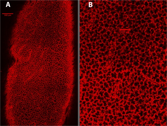
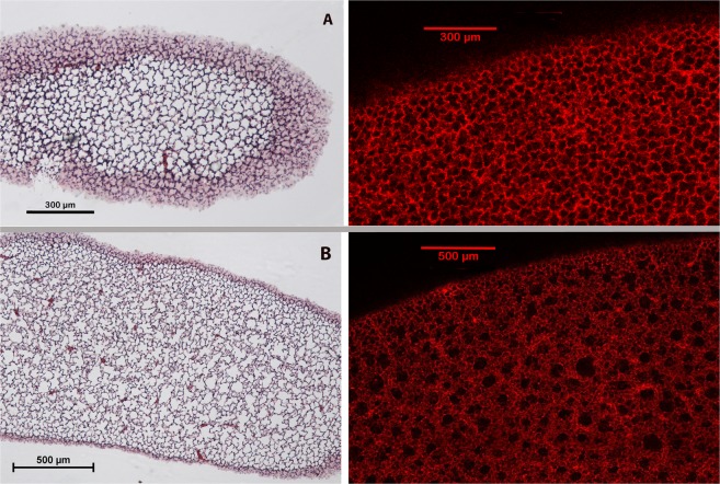
In this study we have utilized an optical clearing method to allow visualization of a heretofore undescribed subpleural acinar structural organization in the mammalian lung. The clearing method enables visualization of the lung structure deep below the visceral pleura in intact inflated lungs. In addition to confirming previous observations that the immediate subpleural alveoli are uniform in appearance, we document for the first time that the subpleural lung parenchyma is much more uniformly organized than the internal parenchyma. Specifically, we report that below the surface layer of alveoli, there is a striking parallel arrangement of alveolar ducts that all run perpendicular to the visceral pleural surface. A three dimensional visualization of alveolar ducts allowed for a calculation of the average inner to outer duct diameter ratio of 0.53 in these subpleural ducts. This unique, self-organizing parallel duct structure likely impacts both elastic recoil and the transmission of tethering forces in healthy and diseased lungs.


Conflict of interest statement

The authors declare no competing interests.

Figures

Figure 1
Figure 1
Low (A) and high (B) magnification images of the subpleural alveoli visualized in a cleared mouse lung at 20 µm below the pleural surface.
Figure 2
Figure 2
Low (A) and high (B) power images of mouse alveolar ducts visualized 100 µm below the visceral pleura. All the black holes in the low power image are duct lumens. (The varying light and dark square regions in the low power image (2A) result from a reconstruction artifact in stitching the separately imaged regional stacks.) In 2(B), where there are adjacent ducts, there is a clear double layer of alveoli between them.
Figure 3
Figure 3
Image showing how a rough estimate of the subpleural duct dimensions was made. Ovals were placed in lumen of circular appearing ducts. A second circle was drawn around the outer border of the alveoli that surround this lumen.
Figure 4
Figure 4
Images from 5 larger species showing the presence of subpleural alveolar ducts as were found in the mice. The rabbit and rat lungs were prepared as in the mouse, but the dog, sheep, and human lungs were not perfused to wash out the blood. For these larger lungs, after the lungs were fixed for at least 2 weeks with formalin, small mouse lung size pieces were cut and placed in the clearing solution. When cleared, they were similarly imaged through the visceral pleural surface. Image depth in the larger species becomes limited very quickly, but in all species, parallel perpendicular ducts are readily seen below the initial subpleural layer of alveoli. Sources of animals: male adult Sprague Dawley rat; male adult NZ White rabbit; male mongrel 20 kg dog; male 25 kg sheep; tissue from 1 human (61 y/o female). The human lung tissue was supplied from another investigator and the lung was not inflated for fixation, as done in all the other species.
Figure 5
Figure 5
Comparison of confocal and histologic images at 20 µm (A) and 80 µm (B) below the visceral pleura. Subpleural alveoli are clearly seen in both the intact and histologic sections at 20 µm, but the duct structure below this initial level is only apparent in the intact lung.

References

    1. Salito C, Aliverti A, Mazzuca E, Rivolta I, Miserocchi G. The effect of exogenous surfactant on alveolar interdependence. Respir. Physiol. Neurobiol. 2015;210:7–13. doi: 10.1016/j.resp.2015.01.009. - DOI - PubMed
    1. Salito C, Aliverti A, Rivolta I, Mazzuca E, Miserocchi G. Alveolar mechanics studied by in vivo microscopy imaging through intact pleural space. Respir. Physiol. Neurobiol. 2014;202:44–49. doi: 10.1016/j.resp.2014.07.012. - DOI - PubMed
    1. Daly BD, Parks GE, Edmonds CH, Hibbs CW, Norman JC. Dynamic alveolar mechanics as studied by videomicroscopy. Respir. Physiol. 1975;24:217–232. doi: 10.1016/0034-5687(75)90115-2. - DOI - PubMed
    1. Mazzuca E, Salito C, Rivolta I, Aliverti A, Miserocchi G. From morphological heterogeneity at alveolar level to the overall mechanical lung behavior: an in vivo microscopic imaging study. Physiological Rep. 2014;2:e00221. doi: 10.1002/phy2.221. - DOI - PMC - PubMed
    1. Moreci AP, Norman JC. Measurements of alveolar sac diameters by incident-light photomicrography. Eff. positive-pressure respiration. Ann. Thorac. Surg. 1973;15:179–186. doi: 10.1016/s0003-4975(10)64951-x. - DOI - PubMed

Publication types