

Kynurenine Pathway as a New Target of Cognitive Impairment Induced by Lead Toxicity During the Lactation
- PMID: 32081969
- PMCID: PMC7035386
- DOI: 10.1038/s41598-020-60159-3
Kynurenine Pathway as a New Target of Cognitive Impairment Induced by Lead Toxicity During the Lactation
Abstract
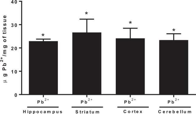
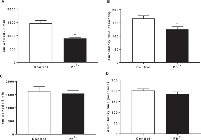
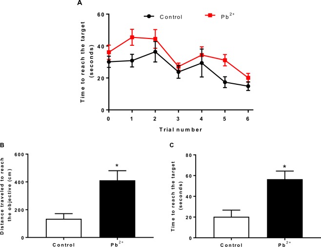
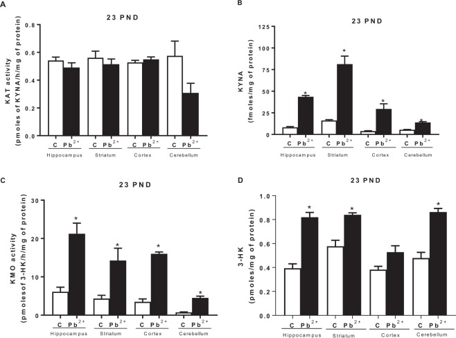
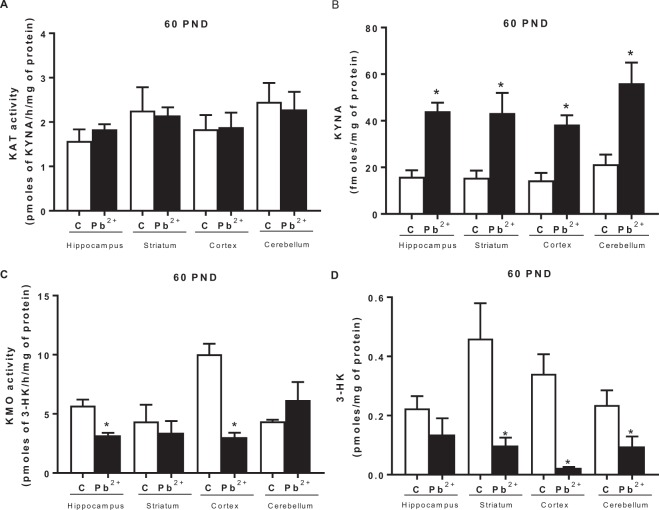
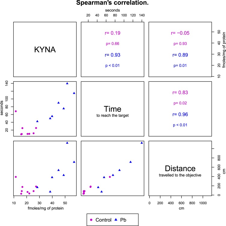
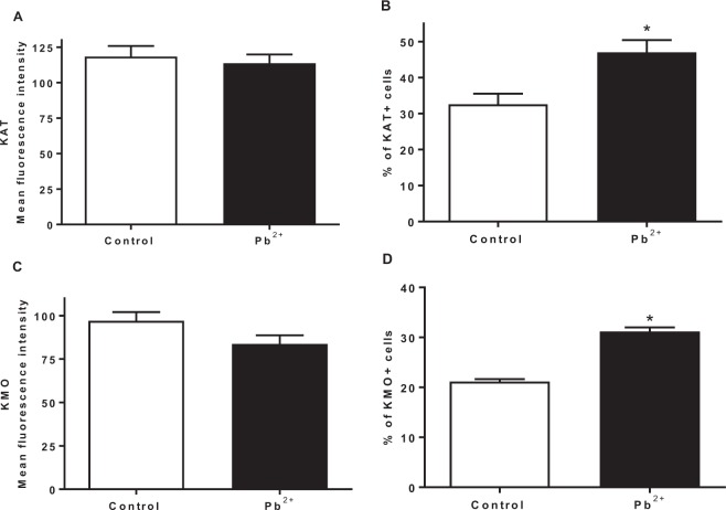
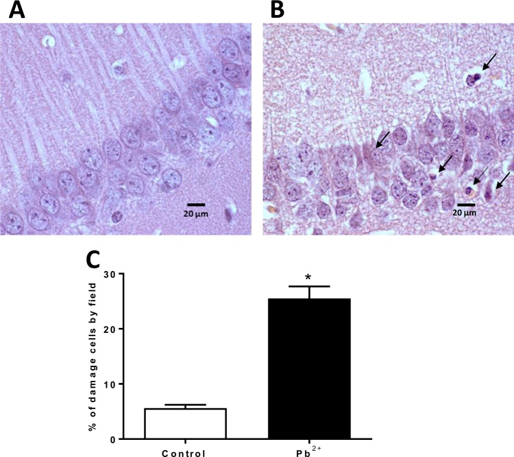
The immature brain is especially vulnerable to lead (Pb2+) toxicity, which is considered an environmental neurotoxin. Pb2+ exposure during development compromises the cognitive and behavioral attributes which persist even later in adulthood, but the mechanisms involved in this effect are still unknown. On the other hand, the kynurenine pathway metabolites are modulators of different receptors and neurotransmitters related to cognition; specifically, high kynurenic acid levels has been involved with cognitive impairment, including deficits in spatial working memory and attention process. The aim of this study was to evaluate the relationship between the neurocognitive impairment induced by Pb2+ toxicity and the kynurenine pathway. The dams were divided in control group and Pb2+ group, which were given tap water or 500 ppm of lead acetate in drinking water ad libitum, respectively, from 0 to 23 postnatal day (PND). The poison was withdrawn, and tap water was given until 60 PND of the progeny. The locomotor activity in open field, redox environment, cellular function, kynurenic acid (KYNA) and 3-hydroxykynurenine (3-HK) levels as well as kynurenine aminotransferase (KAT) and kynurenine monooxygenase (KMO) activities were evaluated at both 23 and 60 PND. Additionally, learning and memory through buried food location test and expression of KAT and KMO, and cellular damage were evaluated at 60 PND. Pb2+ group showed redox environment alterations, cellular dysfunction and KYNA and 3-HK levels increased. No changes were observed in KAT activity. KMO activity increased at 23 PND and decreased at 60 PND. No changes in KAT and KMO expression in control and Pb2+ group were observed, however the number of positive cells expressing KMO and KAT increased in relation to control, which correlated with the loss of neuronal population. Cognitive impairment was observed in Pb2+ group which was correlated with KYNA levels. These results suggest that the increase in KYNA levels could be a mechanism by which Pb2+ induces cognitive impairment in adult mice, hence the modulation of kynurenine pathway represents a potential target to improve behavioural alterations produced by this environmental toxin.
Conflict of interest statement
The authors certify that they have no affiliations with or involvement in any organization or entity with any financial interest (such as honoraria; educational grants; participation in speakers’ bureaus; membership, employment, consultancies, stock ownership, or other equity interest; and expert testimony or patent-licensing arrangements), or non-financial interest (such as personal or professional relationships, affiliations, knowledge or beliefs) in the subject matter or materials discussed in this manuscript.
Figures
Similar articles
-
Modulation of Kynurenic Acid Production by N-acetylcysteine Prevents Cognitive Impairment in Adulthood Induced by Lead Exposure during Lactation in Mice.Antioxidants (Basel). 2023 Nov 23;12(12):2035. doi: 10.3390/antiox12122035. Antioxidants (Basel). 2023. PMID: 38136155 Free PMC article.
-
On the relationship between the two branches of the kynurenine pathway in the rat brain in vivo.J Neurochem. 2009 Apr;109(2):316-25. doi: 10.1111/j.1471-4159.2009.05893.x. Epub 2009 Feb 6. J Neurochem. 2009. PMID: 19226371 Free PMC article.
-
Maternal genotype determines kynurenic acid levels in the fetal brain: Implications for the pathophysiology of schizophrenia.J Psychopharmacol. 2018 Nov;32(11):1223-1232. doi: 10.1177/0269881118805492. Epub 2018 Oct 24. J Psychopharmacol. 2018. PMID: 30354938
-
Galantamine-Memantine Combination and Kynurenine Pathway Enzyme Inhibitors in the Treatment of Neuropsychiatric Disorders.Complex Psychiatry. 2021 Aug;7(1-2):19-33. doi: 10.1159/000515066. Epub 2021 Feb 8. Complex Psychiatry. 2021. PMID: 35141700 Free PMC article. Review.
-
The kynurenine pathway in schizophrenia and bipolar disorder.Neuropharmacology. 2017 Jan;112(Pt B):297-306. doi: 10.1016/j.neuropharm.2016.05.020. Epub 2016 May 28. Neuropharmacology. 2017. PMID: 27245499 Review.
Cited by
-
Exposure to elevated embryonic kynurenine in rats: Sex-dependent learning and memory impairments in adult offspring.Neurobiol Learn Mem. 2020 Oct;174:107282. doi: 10.1016/j.nlm.2020.107282. Epub 2020 Jul 30. Neurobiol Learn Mem. 2020. PMID: 32738461 Free PMC article.
-
Kynurenine and oxidative stress in children having learning disorder with and without attention deficit hyperactivity disorder: possible role and involvement.BMC Neurol. 2022 Sep 20;22(1):356. doi: 10.1186/s12883-022-02886-w. BMC Neurol. 2022. PMID: 36127656 Free PMC article.
-
Early-Life Lead Exposure: Risks and Neurotoxic Consequences.Curr Med Chem. 2024;31(13):1620-1633. doi: 10.2174/0929867330666230409135310. Curr Med Chem. 2024. PMID: 37031386 Review.
-
Evaluation of Analytes Characterized with Potential Protective Action after Rat Exposure to Lead.Molecules. 2021 Apr 9;26(8):2163. doi: 10.3390/molecules26082163. Molecules. 2021. PMID: 33918725 Free PMC article.
-
Kynurenine Monooxygenase Expression and Activity in Human Astrocytomas.Cells. 2021 Aug 8;10(8):2028. doi: 10.3390/cells10082028. Cells. 2021. PMID: 34440798 Free PMC article.
References
-
- Garza A, Vega R, Soto E. Cellular mechanisms of lead neurotoxicity. Med. Sci. Monit. 2006;12:RA57–65. - PubMed
Publication types
MeSH terms
Substances
LinkOut - more resources
Full Text Sources
Miscellaneous
