

Thymic Epithelial Cells Contribute to Thymopoiesis and T Cell Development
- PMID: 32082299
- PMCID: PMC7005006
- DOI: 10.3389/fimmu.2019.03099
Thymic Epithelial Cells Contribute to Thymopoiesis and T Cell Development
Erratum in
-
Corrigendum: Thymic Epithelial Cells Contribute to Thymopoiesis and T Cell Development.Front Immunol. 2020 Nov 30;11:628464. doi: 10.3389/fimmu.2020.628464. eCollection 2020. Front Immunol. 2020. PMID: 33329618 Free PMC article.
Abstract
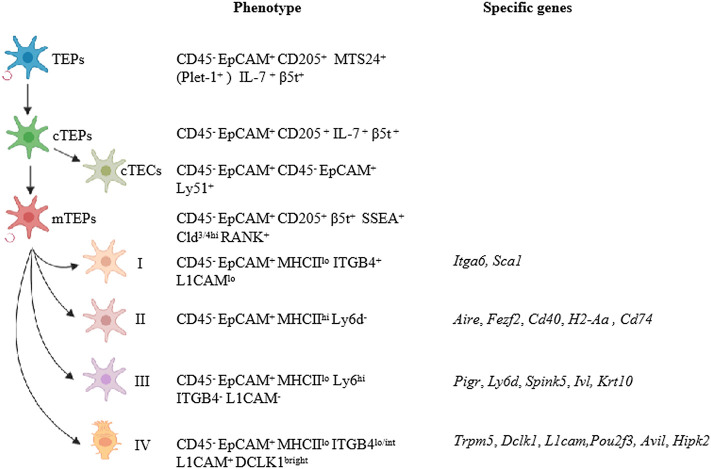
The thymus is the primary lymphoid organ responsible for the generation and maturation of T cells. Thymic epithelial cells (TECs) account for the majority of thymic stromal components. They are further divided into cortical and medullary TECs based on their localization within the thymus and are involved in positive and negative selection, respectively. Establishment of self-tolerance in the thymus depends on promiscuous gene expression (pGE) of tissue-restricted antigens (TRAs) by TECs. Such pGE is co-controlled by the autoimmune regulator (Aire) and forebrain embryonic zinc fingerlike protein 2 (Fezf2). Over the past two decades, research has found that TECs contribute greatly to thymopoiesis and T cell development. In turn, signals from T cells regulate the differentiation and maturation of TECs. Several signaling pathways essential for the development and maturation of TECs have been discovered. New technology and animal models have provided important observations on TEC differentiation, development, and thymopoiesis. In this review, we will discuss recent advances in classification, development, and maintenance of TECs and mechanisms that control TEC functions during thymic involution and central tolerance.
Keywords: medullary thymic epithelial cells (mTECs); thymic epithelial cells (TECs); thymopoiesis; tissue-restricted antigens (TRAs); tolerance.
Copyright © 2020 Wang, Pan, Zheng, Zhong, Tan, Liang, He, Feng, Zhao and Qiu.
Figures
References
Publication types
MeSH terms
Substances
LinkOut - more resources
Full Text Sources
Miscellaneous