. 2020 Jan 31:10:1409.
doi: 10.3389/fgene.2019.01409. eCollection 2019.

LncRNA LUCRC Regulates Colorectal Cancer Cell Growth and Tumorigenesis by Targeting Endoplasmic Reticulum Stress Response

Affiliations

LncRNA LUCRC Regulates Colorectal Cancer Cell Growth and Tumorigenesis by Targeting Endoplasmic Reticulum Stress Response

Guo-Hui Tang et al. Front Genet. .

Abstract

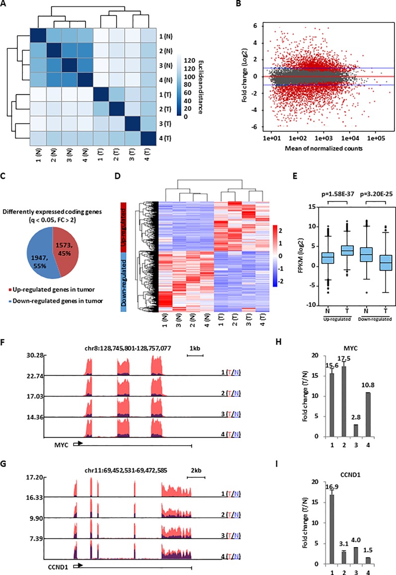
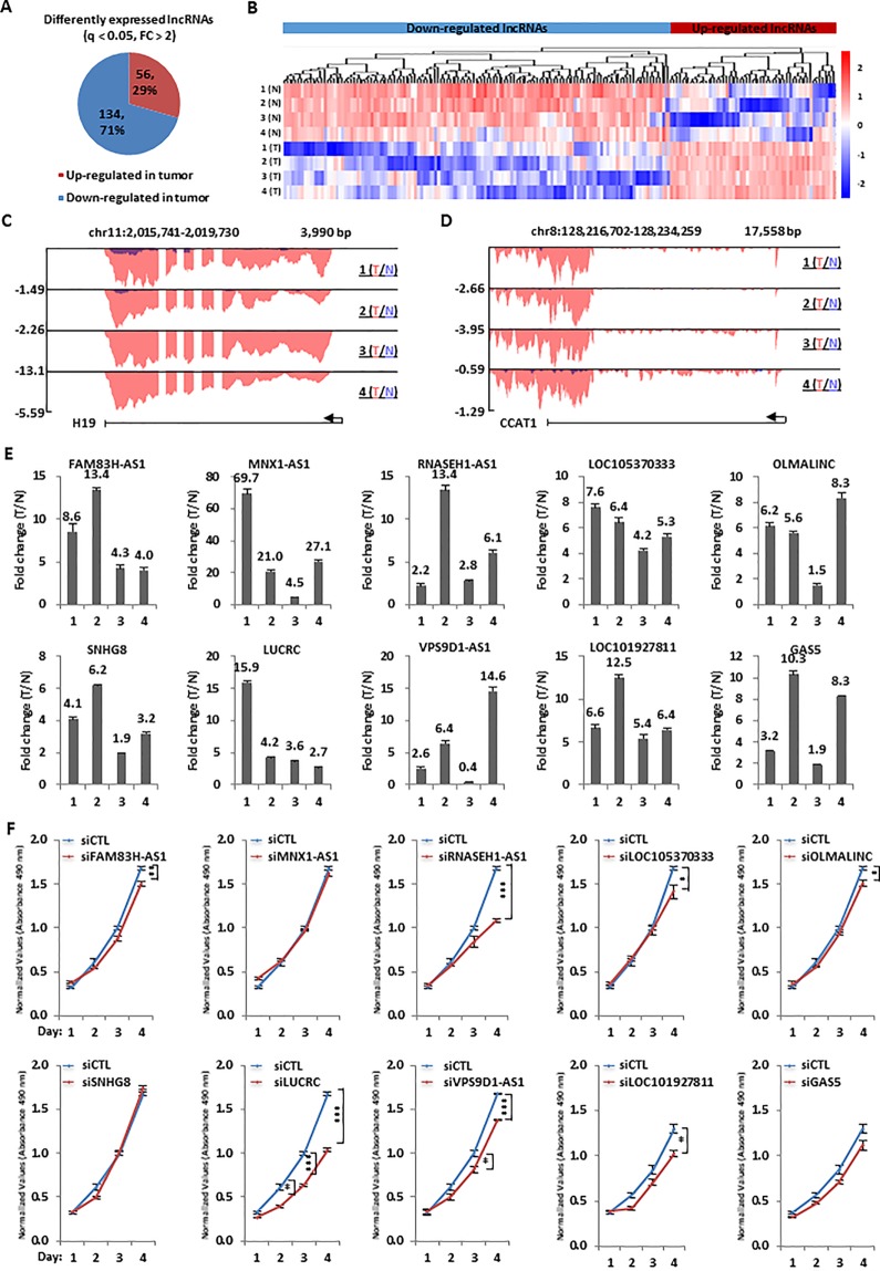
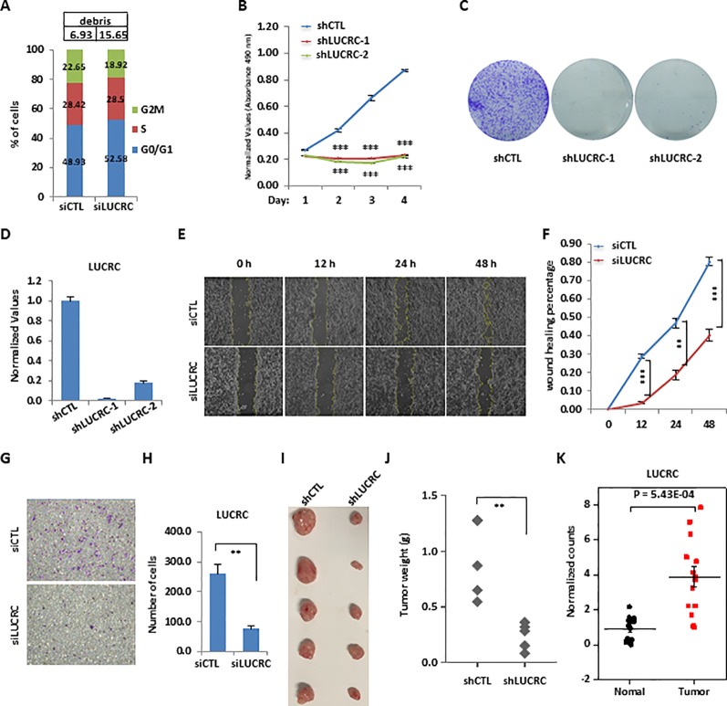
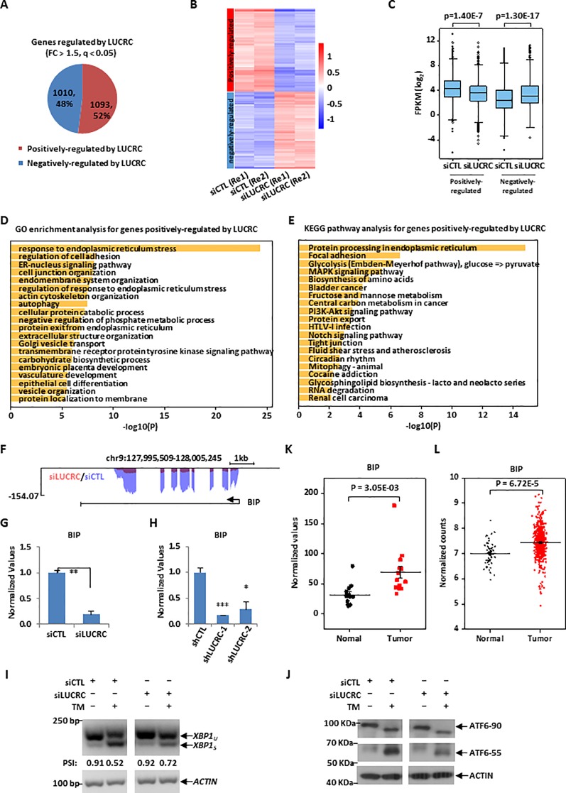
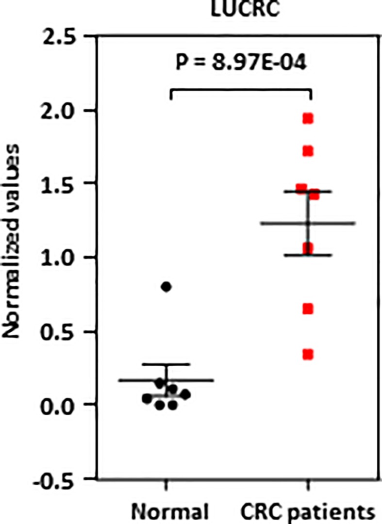
Colorectal cancer (CRC) is the second most common cause of cancer-related death worldwide, and is well known for its strong invasiveness, rapid recurrence, and poor prognosis. Long non-coding RNAs (lncRNAs) have been shown to be involved in the development of various types of cancers, including colorectal cancer. Here, through transcriptomic analysis and functional screening, we reported that lncRNA LUCRC (LncRNA Upregulated in Colorectal Cancer) is highly expressed in colorectal tumor samples and is required for colorectal cancer cell proliferation, migration, and invasion in cultured cells and tumorigenesis in xenografts. LUCRC was found to regulate target gene expression of unfolded protein response (UPR) in endoplasmic reticulum (ER), such as BIP. The clinical significance of LUCRC is underscored by the specific presence of LUCRC in blood plasma of patients with colorectal cancers. These findings revealed a critical regulator of colorectal cancer development, which might serve as a therapeutic target in colorectal cancer.

Keywords: cell growth; colorectal cancer; long non-coding RNA; therapeutic target; unfolded protein response.


Figures

Figure 1
Figure 1
A large cohort of genes were dysregulated in colorectal cancer. (A) Colorectal tumor tissues (T) and the corresponding adjacent normal tissues (N) (n = 4 pairs) were collected and subjected to RNA-seq analysis followed by hierarchical cluster analysis. (B) MA plot shows the fold change (FC, tumor/normal, log2) against the average of normalized counts for all the samples as described in (A). Red dots represented genes with significant change in tumor tissues (q <0.05), and blue line indicated fold change of two. (C) Pie chart shows genes dysregulated, both up-regulated and down-regulated, in colorectal tumor samples as described in (A) (q < 0.05, FC > 2). (D, E) Heat map (D) and box plot (E) representation of the expression levels (FPKM, log2) for genes, both up-regulated and down-regulated, in colorectal tumor samples as described in (C). (F, G) UCSC genome browser views of RNA-seq as described in (A) for specific genes were shown as indicated. (H, I) RNA samples from four colorectal cancer patients as described in (A) were subjected to RT-qPCR analysis to examine the expression of MYC (H) and CCND1 (I). Data was presented as fold change of tumor (T) versus normal (N) as indicated (± s.e.m.).
Figure 2
Figure 2
Hundreds of lncRNAs were dysregulated and a number of them were required for colorectal cancer cell growth. (A) Pie chart shows lncRNAs dysregulated, both up-regulated and down-regulated, in colorectal tumor samples as described in Figure 1A (q < 0.05, FC > 2). (B) Heat map representation of the expression levels (FPKM, log2) for dysregulated lncRNAs, both up-regulated and down-regulated, in colorectal tumor samples as described in (A). (C, D) UCSC genome browser views of RNA-seq as described in Figure 1A for specific up-regulated lncRNAs were shown as indicated. (E) RNA samples from four patients with colorectal cancer as described in Figure 1A were subjected to RT-qPCR analysis to examine the expression of 10 top-upregulated lncRNAs in colorectal tumor samples as detected by RNA-seq analysis. Data was presented as fold change of tumor (T) versus normal (N) as indicated (± s.e.m.). (F) HCT116 cells were transfected with control siRNA (siCTL) or siRNA specifically targeting each individual lncRNA for duration as indicated followed by cell proliferation assay. (± s.e.m., *P < 0.05, **P < 0.01, ***P < 0.001).
Figure 3
Figure 3
LUCRC was required for colorectal cancer cell proliferation, migration, and invasion in vitro and tumorigenesis in vivo. (A) HCT116 cells were transfected with control siRNA (siCTL) or siRNA specifically against (siLUCRC) for 3 days before FACS analysis. Percentage of apoptotic cells (debris) were also shown on the top. (B) HCT116 cells were infected with control shRNA (shCTL) or two independent shRNA specifically targeting LUCRC (shLUCRC) for duration as indicated before cell proliferation assay (± s.e.m., ***P < 0.001). (C) HCT116 cells were infected with shCTL or two independent shLUCRC for 10 days before colony formation assay (± s.e.m., **P < 0.01, ***P < 0.001). (D) HCT116 cells as described in (B, C) were subjected to RNA extraction and RT-qPCR analysis to examined the expression of LUCRC (± s.e.m., ***P < 0.001). (E) HCT116 cells were transfected with siCTL or siLUCRC for 2 days and then re-seeded at full confluence and maintained for duration as indicated before wound-healing assay. (F) Quantification of wound closure as shown in (E) (± s.e.m., **P < 0.01, ***P < 0.001). (G) HCT116 cells were transfected with siCTL or siLUCRC for 2 days and then re-seeded at full confluence and maintained for 1 day before transwell assay. (H) Quantification of cells as shown in (G) (± s.e.m., **P < 0.01). (I) ShCTL or shLUCRC-infected HCT116 cells were injected subcutaneously into female BALB/C nude mice for xenograft experiments. (J) Tumor weight as shown in (I) (± s.e.m., **P < 0.01). (K) Colorectal tumor tissues and the corresponding adjacent normal tissues were collected from a group of colorectal cancer patients (n = 14) and subjected to RNA extraction and RT-qPCR analysis to examine the expression of LUCRC (± s.e.m., ***P < 0.001).
Figure 4
Figure 4
LUCRC was required for the expression of genes involved in ER stress response, including BIP. (A) HCT116 cells transfected with control siRNA (siCTL) and siRNA specifically targeting LUCRC (siLUCRC) for 3 days were subjected to RNA-seq analysis, and genes positively- and negatively-regulated by LUCRC were shown by pie chart (Fold change (FC) > 1.5). (B, C) Heat map (B) and box plot (C) representation of the expression levels (FPKM, log2) for genes positively- and negatively-regulated by LUCRC in HCT116 cells as shown in (A). Re1: replicate 1; Re2: replicate 2. (D, E) GO (D) and KEGG (E) analysis for genes positively-regulated by LUCRC in HCT116 cells as shown in (A). (F) UCSC genome browser view of RNA-seq as described in (A) for BIP was shown. (G) HCT116 cells were transfected with siCTL or siLUCRC for 3 days followed by RT-qPCR analysis to examine the expression of BIP (± s.e.m., **P < 0.01). (H) HCT116 cells were infected with shCTL or shLUCRC for 3 days followed by RT-qPCR analysis to examine the expression of BIP (± s.e.m., *P < 0.05, ***P < 0.001). (I) HCT116 cells were transfected with siCTL or siLUCRC for 3 days and then treated with or without tunicamycin (TM) (1μg/mL) for 8 h, followed by RNA extraction, reverse transcription and PCR analysis using primers targeting XBP1 or ACTIN. Splicing of XBP1 was presented as PSI (percentage of inclusion: XBP1u/(XBP1u + XBPIs)). XBP1u: unspliced XBP1; XBP1s: spliced XBP1. DNA fragment size was indicated on the left. bp: base pair. (J) HCT116 cells as described in (I) were subjected to immunoblotting analysis by using antibodies as indicated. Molecular weight was indicated on the left. KDa: kilodalton. ATF6-90: full length ATF6; ATF6-55: partial ATF6. (K) Colorectal tumor tissues and the corresponding adjacent normal tissues were collected from a group of colorectal cancer patients (n = 14) and subjected to RNA extraction and RT-qPCR analysis to examine the expression of BIP (± s.e.m., **P < 0.01). (L) The expression of BIP in a cohort of clinical colorectal tumor (n = 647) and normal (n = 51) samples from TCGA (The Cancer Genome Atlas).
Figure 5
Figure 5
LUCRC was detectable in blood samples of colorectal cancer patients. Blood samples from seven colorectal cancer patients as well as seven healthy controls were subjected to RNA extraction and RT-qPCR analysis.

References

    1. Anderson D. M., Anderson K. M., Chang C. L., Makarewich C. A., Nelson B.R., McAnally J.R., et al. (2015). A micropeptide encoded by a putative long noncoding RNA regulates muscle performance. Cell 160, 595–606. 10.1016/j.cell.2015.01.009 - DOI - PMC - PubMed
    1. Bian Z., Jin L., Zhang J., Yin Y., Quan C., Hu Y., et al. (2016). LncRNA-UCA1 enhances cell proliferation and 5-fluorouracil resistance in colorectal cancer by inhibiting miR-204-5p. Sci. Rep. 6, 23892. 10.1038/srep23892 - DOI - PMC - PubMed
    1. Board P.D.Q.A.T.E (2002). “Colon Cancer Treatment (PDQ(R)): Health Professional Version,” in PDQ Cancer Information Summaries ((US), Bethesda (MD);: National Cancer Institute; ).
    1. Bouckenheimer J., Assou S., Riquier S., Hou C., Philippe N., Sansac C., et al. (2016). Long non-coding RNAs in human early embryonic development and their potential in ART. Hum. Reprod. Update 23, 19–40. 10.1093/humupd/dmw035 - DOI - PubMed
    1. Bray F., Ferlay J., Soerjomataram I., Siegel R.L., Torre L.A., Jemal A., et al. (2018). Global cancer statistics 2018: GLOBOCAN estimates of incidence and mortality worldwide for 36 cancers in 185 countries. CA Cancer J. Clin. 68, 394–424. 10.3322/caac.21492 - DOI - PubMed