. 2020 Feb 13:8:6.
doi: 10.1186/s40364-020-0185-8. eCollection 2020.

Increased PD-1+Tim-3+ exhausted T cells in bone marrow may influence the clinical outcome of patients with AML

Affiliations

Increased PD-1+Tim-3+ exhausted T cells in bone marrow may influence the clinical outcome of patients with AML

Jiaxiong Tan et al. Biomark Res. .

Abstract

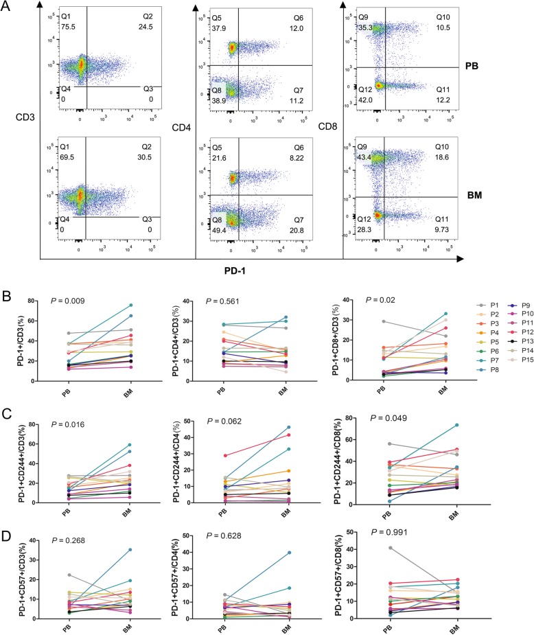
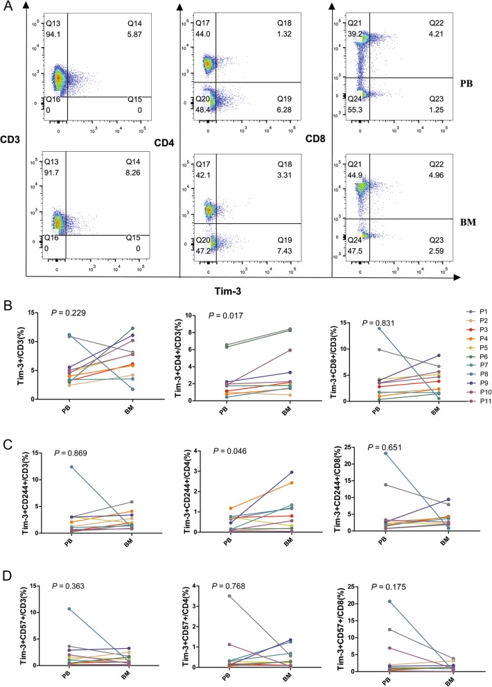
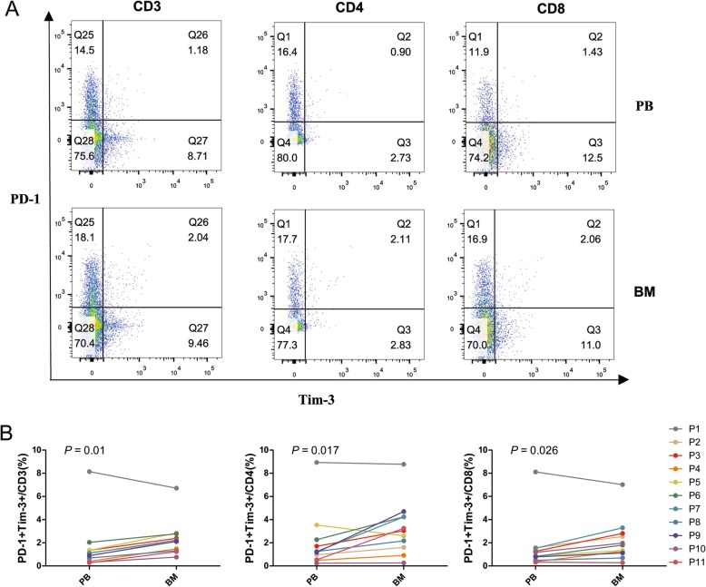
Background: Altered expression of T cell immune inhibitory receptors may result in immunosuppression and associate with the poor prognosis of leukemia patients in which the leukemic bone marrow (BM) microenvironment may contribute to such immunosuppression. We found higher numbers of programmed death-1 (PD-1) + exhausted T cells in peripheral blood (PB) from acute myeloid leukemia (AML) patients. To investigate the leukemic BM influence on immunosuppression, we further compared the distributions of PD-1 and T cell immunoglobulin mucin-3 (Tim-3) and the exhausted T cell phenotype in PB and BM from AML patients and characterized their relationship with clinical outcome.

Methods: PB and BM samples from 15 patients with newly diagnosed AML were collected and analyzed for the expression of PD-1, Tim-3, CD244, and CD57 on CD3+, CD4+, and CD8+ T cells by multicolor flow cytometry.

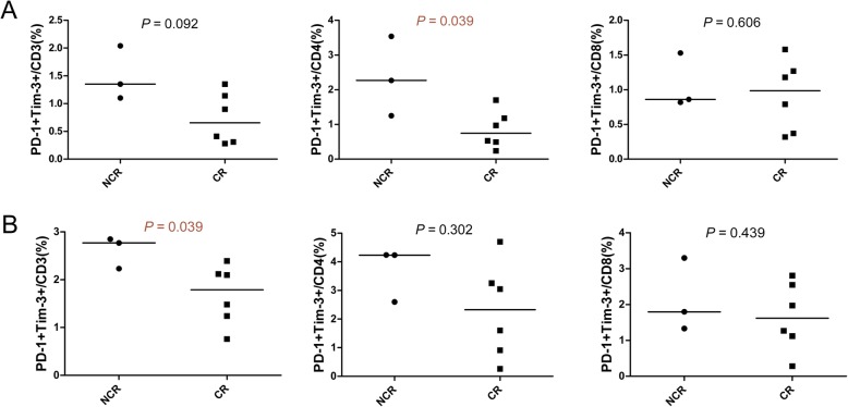
Results: The proportions of PD-1 + CD3+ and PD-1 + CD8+ T cells were significantly higher in BM compared with PB. Similarly, higher PD-1 + CD244 + CD3+ and PD-1 + CD244 + CD8+ T cells were found in BM, and an increased tendency for PD-1 + CD244 + CD4+ T cells was also detected in this group. In contrast, increased Tim-3 + CD4+/Tim-3 + CD244 + CD4+ T cells were predominant in BM compared with PB, but there was no statistically significant difference in Tim-3 + CD8+ T cells. Moreover, PD-1 and Tim-3 double-positive CD3+/CD4+/CD8+ T cells were significantly increased in the BM group. In addition, a higher proportion of PD-1 + Tim-3 + CD3+ T cells in the BM and PD-1 + Tim-3 + CD4+ T cells in PB was detected in non-complete remission (NCR) compared with complete remission (CR) patients after first-cycle chemotherapy.

Conclusions: Upregulation of PD-1 and Tim-3 and the exhausted phenotype of CD4+ and CD8+ T cells in the BM of AML patients may contribute to mediating the leukemic immunosuppressive microenvironment, and increased PD-1 + Tim-3+ CD8+ T cells may be related to T cell dysfunction in AML, which may influence clinical outcome.

Keywords: AML; PD-1; T cell exhaustion; T cell subset; Tim-3.


Conflict of interest statement

Competing interestsThe authors declare that they have no competing interests.

Figures

Fig. 1
Fig. 1
Distribution of PD-1+, CD57+, CD244+ T cells in the CD3+, CD4+ and CD8+ T cell subsets in PB or BM from patients with AML. A: Flow cytometry detection of PD-1 + CD3+, PD-1 + CD4+, and PD-1 + CD8+ T cells in PB and BM in a patient with AML. B: Comparison of the percentage of PD-1 + CD3+, PD-1 + CD4+, PD-1 + CD8+ T cells in PB and BM from 15 patients (P1 to P15) with AML. C: Comparison of the percentage of PD-1 + CD244 + CD3+, PD-1 + CD244 + CD4+, and PD-1 + CD244 + CD8+ T cells in PB and BM from 15 patients with AML. D: Comparison of the percentage of PD-1 + CD57 + CD3+, PD-1 + CD57 + CD4+, and PD1 + CD57 + CD8+ T cells in PB and BM from 15 patients with AML
Fig. 2
Fig. 2
Distribution of Tim-1+, CD57+, CD244+ T cells in CD3+, CD4+ and CD8+ T cell subsets in PB and BM from patients with AML. A: Flow cytometry detection of Tim-3 + CD3+, Tim-3 + CD4+, and Tim-3 + CD8+ T cells in PB and BM from one patient with AML. B: Comparison of the percentage of Tim-3 + CD3+, Tim-3 + CD4+, Tim-3 + CD8+ T cells in PB and BM from 11 patients (P1 to P11) with AML. C: Comparison of the percentage of Tim-3 + CD244 + CD3+, Tim-3 + CD244 + CD4+, and Tim-3 + CD244 + CD8+ T cells in PB and BM from 11 patients with AML. D: Comparison of the percentage of Tim-3 + CD57 + CD3+, Tim-3 + CD57 + CD4+, and Tim-3 + CD57 + CD8+ T cells in PB and BM from 11 patients with AML
Fig. 3
Fig. 3
Distribution of PD-1 + Tim-1 + T cells in the CD3+, CD4+, and CD8+ T cell subsets in PB and BM from patients with AML. A: Flow cytometry detection of PD-1 + Tim-3 + CD3+, CD4+ and CD8+ T cells in PB and BM from one patient with AML. B: Comparison of the percentage of PD-1 + Tim-3 + CD3+, CD4+, and CD8+ T cells in PB and BM from 11 patients (P1 to P11) with AML by flow cytometry analysis
Fig. 4
Fig. 4
Differences in the distribution of PD-1+ T cells in BM and PB between patients with complete remission and those with non-complete remission after first-cycle chemotherapy as measured by flow cytometry analysis. A: Comparison of the percentage of PD-1 + Tim-3 + CD3+, CD4+, and CD8+ T cells in PB from patients in complete remission (CR, n = 6) and non-complete remission (NCR, n = 3). B: Comparison of the percentage of PD-1 + Tim-3 + CD3+, CD4+, and CD8+ T cells in BM from patients in the CR and NCR groups

References

    1. Beyar-Katz O, Gill S. Novel approaches to acute myeloid leukemia immunotherapy. Clin Cancer Res. 2018;24(22):5502–5515. doi: 10.1158/1078-0432.CCR-17-3016. - DOI - PubMed
    1. Wang M, Bu J, Zhou M, Sido J, Lin Y, Liu G, et al. CD8(+)T cells expressing both PD-1 and TIGIT but not CD226 are dysfunctional in acute myeloid leukemia (AML) patients. Clin Immunol. 2018;190:64–73. doi: 10.1016/j.clim.2017.08.021. - DOI - PubMed
    1. Yang X, Wang J. Precision therapy for acute myeloid leukemia. J Hematol Oncol. 2018;11(1):3. doi: 10.1186/s13045-017-0543-7. - DOI - PMC - PubMed
    1. Chisini M, Stefanizzi C, Ceglie T, Raponi S, Vozella F, Colafigli G, et al. Independent prognostic impact of CD15 on complete remission achievement in patients with acute myeloid leukemia. Hematol Oncol. 2017;35(4):804–809. doi: 10.1002/hon.2331. - DOI - PubMed
    1. Yu B, Liu D. Gemtuzumab ozogamicin and novel antibody-drug conjugates in clinical trials for acute myeloid leukemia. Biomark Res. 2019;7:24. doi: 10.1186/s40364-019-0175-x. - DOI - PMC - PubMed