Case Reports
. 2020 Jan 28:2020:5492754.
doi: 10.1155/2020/5492754. eCollection 2020.

Primary Intraosseous Synovial Sarcoma with Molecular Confirmation: Expanding and Clarifying the Spectrum of This Rare Neoplasm

Affiliations
Case Reports

Primary Intraosseous Synovial Sarcoma with Molecular Confirmation: Expanding and Clarifying the Spectrum of This Rare Neoplasm

Kelsey E McHugh et al. Case Rep Pathol. .

Abstract

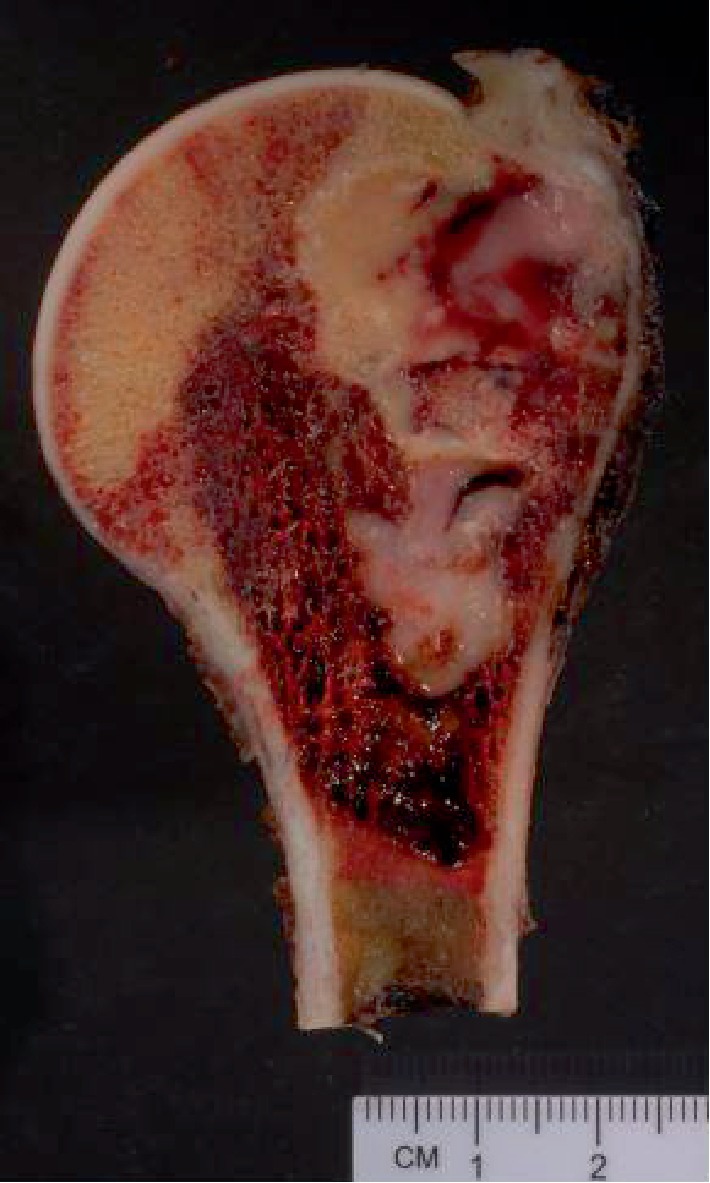
Synovial sarcoma is a well-known malignant tumor usually originating within deep soft tissues of the lower extremities of adolescents and young adults. Rare radiologically confirmed examples of primary bone synovial sarcoma have been documented, generally in isolated case reports. Herein, we report two cases of primary intraosseous synovial sarcoma, with molecular confirmation, involving the left humerus of a 45-year-old female and the right fourth metatarsal bone in a 36-year-old male. Additionally, we clarify the spectrum of primary intraosseous synovial sarcoma by separately analyzing reported cases with radiographic confirmation of bone origin and molecular support for the diagnosis. There are clinicopathologic differences between those tumors with documented molecular confirmation and those lacking such confirmation, specifically regarding their anatomic distribution (p < 0.0001). Regarding the radiology of our two cases, the humeral lesion appeared almost entirely intramedullary without soft tissue extension; the midfoot lesion demonstrated a destructive, metatarsal-centered bone lesion, initially thought clinically to represent primary bone osteosarcoma. The diagnoses of monophasic synovial sarcoma were rendered via core needle biopsies, with molecular FISH confirmation of SYT gene rearrangement. Clinical follow-up data was only available for the female patient with the primary humeral lesion, who underwent surgical resection, with no local recurrence or distant metastasis at 7 months postsurgery. To our knowledge, these are the first reported examples of molecularly confirmed, primary intraosseous synovial sarcomas of the humerus and metatarsal bones. Primary intraosseous synovial sarcomas with molecular confirmation differ clinically from those lacking it; however, the demographic features and metastatic potential appear similar to primary soft tissue synovial sarcoma.


Conflict of interest statement

The authors declare that there are no conflicts of interest regarding the publication of this paper.

Figures

Figure 1
Figure 1
Case 1: proximal left humerus resection showing a fleshy tan-gray lesion involving the intramedullary space with destruction of overlying cortical bone and associated foci of hemorrhage and necrosis.
Figure 2
Figure 2
Case 1: sagittal left shoulder MRI showing a 6 cm intramedullary lesion involving the proximal left humerus with associated periostitis. The lesion was hypointense on T1-weighted images (a) and hyperintense on T2-weighted images (b).
Figure 3
Figure 3
Case 1: resection specimen demonstrating cortical erosion with bony permeation (a). Scattered foci of hemangiopericytoma-like vasculature were present (b). The tumor was monophasic, comprised of bland, ovoid spindle cells arranged in storiform (c) and fascicular (d) patterns.
Figure 4
Figure 4
Case 1: H&E-stained core needle biopsy material (a) and associated immunohistochemistry results demonstrating diffuse membranous positivity for CD99 (b) and focal positivity for EMA (c). CD34 highlights the vasculature of the tumor and is negative in tumor cells (d). The tumor cells are also negative for pankeratin AE1/3 (e) and STAT6 (f).
Figure 5
Figure 5
Cases 1 and 2: positive fluorescence in situ hybridization (FISH) analysis for SYT (SS18) gene rearrangement, performed at the Cleveland Clinic, demonstrated by an abnormal signal pattern seen as disruption of the SYT gene through the breaking apart of the red and green probe signals. In case 2, SYT gene rearrangement was seen in 94% of tumor nuclei (a). In case 1, SYT gene rearrangement was seen in 76% of tumor nuclei (b).
Figure 6
Figure 6
Case 2: anteroposterior left foot radiograph showing a lytic and destructive mass centered on the 4th metatarsal, with destructive extension to the 3rd metatarsal and adjacent cuneiform bones. Punctate calcifications consistent with foci of mineralization can be seen throughout the lesion.
Figure 7
Figure 7
Case 2: core needle biopsy with permeation of spindle cells around bony trabeculae (a). Hemangiopericytoma-like vasculature was identifiable (b). The tumor was monophasic, comprised of uniform, ovoid spindle cells with scanty cytoplasm arranged in short fascicles (c, d).
Figure 8
Figure 8
Case 2: H&E-stained core needle biopsy material (a) and associated immunohistochemistry results demonstrating strong diffuse cytoplasmic positivity for vimentin (b) and membranous positivity for CD99 (c). CD34 highlights the vasculature of the tumor and is negative in tumor cells (d). The tumor cells are also negative for pankeratin (e) and STAT6 (f).

References

    1. Goldblum J. R., Weiss S. W., Folpe A. L. Enzinger and Weiss’s Soft Tissue Tumors. Saunders; 2013. Synovial sarcoma; pp. P1052–P1070.
    1. Cohen I. J., Issakov J., Avigad S., et al. Synovial sarcoma of bone delineated by spectral karyotyping. The Lancet. 1997;350(9092):1679–1680. doi: 10.1016/S0140-6736(05)64278-X. - DOI - PubMed
    1. Jung S. C., Choi J. A., Chung J. H., Oh J. H., Lee J. W., Kang H. S. Synovial sarcoma of primary bone origin: a rare case in a rare site with atypical features. Skeletal Radiology. 2007;36(1):67–71. doi: 10.1007/s00256-006-0185-2. - DOI - PubMed
    1. Beck S. E., Nielsen G. P., Raskin K. A., Schwab J. H. Intraosseous synovial sarcoma of the proximal tibia. International Journal of Surgical Oncology. 2011;2011:5. doi: 10.1155/2011/184891.184891 - DOI - PMC - PubMed
    1. Horvai A., Dashti N. K., Rubin B. P., et al. Genetic and molecular reappraisal of spindle cell adamantinoma of bone reveals a small subset of misclassified intraosseous synovial sarcoma. Modern Pathology. 2019;32(2):231–241. doi: 10.1038/s41379-018-0115-6. - DOI - PubMed

Publication types

LinkOut - more resources