

Rare operations in pediatric heart surgery: Cardiac tumors in childhood
- PMID: 32082795
- PMCID: PMC7018197
- DOI: 10.5606/tgkdc.dergisi.2018.16147
Rare operations in pediatric heart surgery: Cardiac tumors in childhood
Abstract
Background: In this study, we present our 12-year experience in the surgical treatment of primary cardiac tumors in childhood.
Methods: Thirteen pediatric patients (8 males, 5 females; mean age 1.3±1.9 years; range, 3 days to 6 years) who were operated for a primary cardiac tumor in our center between January 2005 and December 2017 were included in this study. The data were evaluated retrospectively based on our medical records.
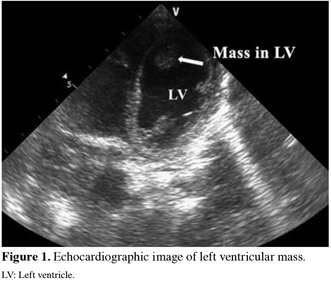
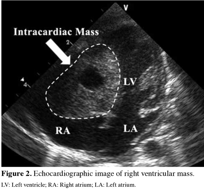
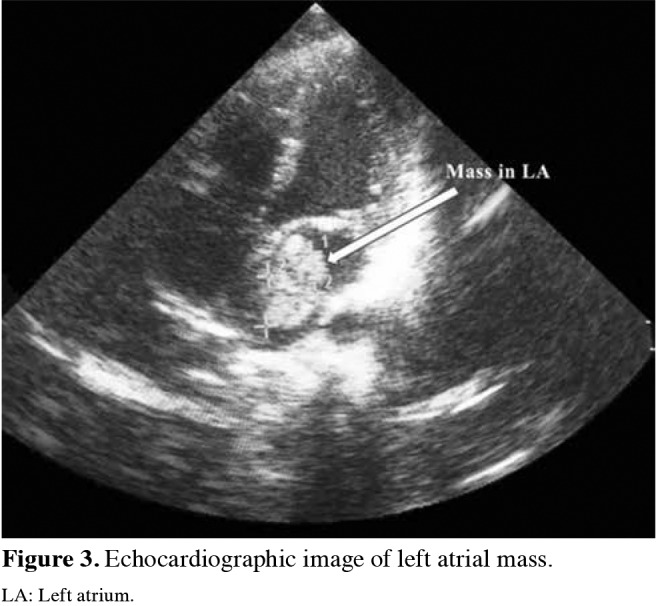
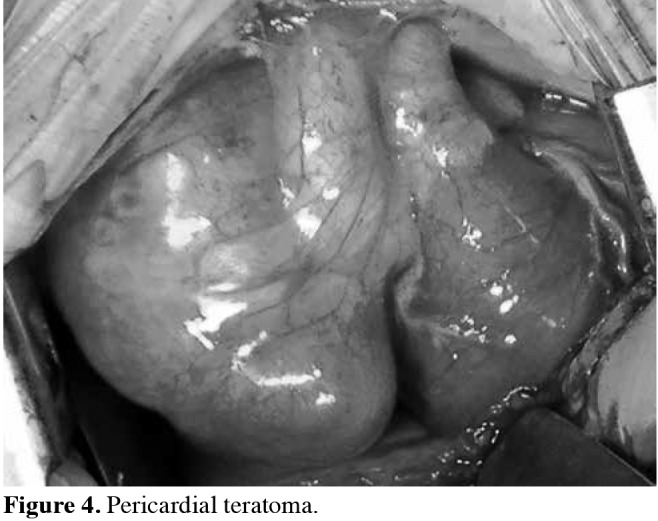
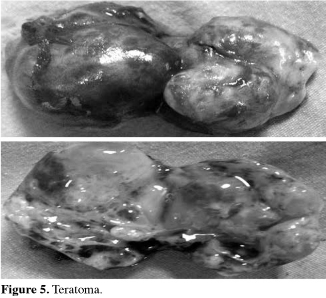
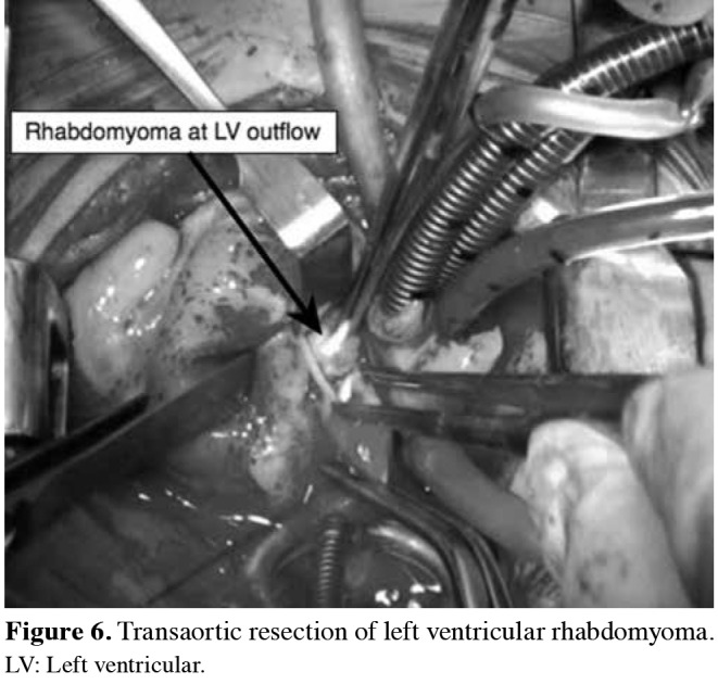
Results: All of the masses resected were benign. However, the most common tumor was rhabdomyoma (n=7), followed by fibroma (n=3), myxoma (n=2), and pericardial teratoma (n=1). The mortality rate was 15.4%, as two patients died in the early postoperative period. No residual mass or tumor recurrence was observed in the early and late postoperative period in the remaining patients.
Conclusion: Although primary cardiac tumors in childhood are usually benign, they may cause clinically significant problems depending on the localization and size of the tumor. Surgical tumor excision is often associated with good long-term outcomes.
Keywords: Childhood; heart surgery; primary cardiac tumors.
Copyright © 2018, Turkish Society of Cardiovascular Surgery.
Conflict of interest statement
Conflict of Interest: The authors declared no conflicts of interest with respect to the authorship and/or publication of this article.
Figures
Similar articles
-
Identification and clinical course of 166 pediatric cardiac tumors.Eur J Pediatr. 2017 Feb;176(2):253-260. doi: 10.1007/s00431-016-2833-4. Epub 2017 Jan 10. Eur J Pediatr. 2017. PMID: 28074279
-
Early and late results in surgical excision of primary cardiac tumors: Our single-institution experience.Turk Gogus Kalp Damar Cerrahisi Derg. 2018 Apr 30;26(2):177-182. doi: 10.5606/tgkdc.dergisi.2018.14985. eCollection 2018 Apr. Turk Gogus Kalp Damar Cerrahisi Derg. 2018. PMID: 32082732 Free PMC article.
-
Pediatric cardiac tumors: a 45-year, single-institution review.World J Pediatr Congenit Heart Surg. 2015 Apr;6(2):215-9. doi: 10.1177/2150135114563938. World J Pediatr Congenit Heart Surg. 2015. PMID: 25870340
-
Primary heart tumors in the pediatric age group: a review of salient pathologic features relevant for clinicians.Pediatr Cardiol. 2000 Jul-Aug;21(4):317-23. doi: 10.1007/s002460010071. Pediatr Cardiol. 2000. PMID: 10865004 Review.
-
Primary cardiac and pericardial neoplasms: radiologic-pathologic correlation.Radiographics. 2000 Jul-Aug;20(4):1073-103; quiz 1110-1, 1112. doi: 10.1148/radiographics.20.4.g00jl081073. Radiographics. 2000. PMID: 10903697 Review.
Cited by
-
Cardiac Myosarcoma in a Newborn Infant-A Case Report and Literature Review.Front Cardiovasc Med. 2021 Jul 14;8:675202. doi: 10.3389/fcvm.2021.675202. eCollection 2021. Front Cardiovasc Med. 2021. PMID: 34336944 Free PMC article.
References
-
- Patel J, Sheppard MN. Pathological study of primary cardiac and pericardial tumours in a specialist UK Centre: surgical and autopsy series. Cardiovasc Pathol. 2010;19:343–352. - PubMed
-
- Motwani M, Kidambi A, Herzog BA, Uddin A, Greenwood JP, Plein S. MR imaging of cardiac tumors and masses: a review of methods and clinical applications. Radiology. 2013;268:26–43. - PubMed
-
- Kutsal A, Koç M. Left ventricular myxoma. EJCM. 2015;3:27–30.
-
- Barreiro M, Renilla A, Jimenez JM, Martin M, Al Musa T, Garcia L, et al. Primary cardiac tumors: 32 years of experience from a Spanish tertiary surgical center. Cardiovasc Pathol. 2013;22:424–427. - PubMed
-
- Beghetti M, Gow RM, Haney I, Mawson J, Williams WG, Freedom RM. Pediatric primary benign cardiac tumors: a 15-year review. Am Heart J. 1997;134:1107–1114. - PubMed
LinkOut - more resources
Full Text Sources