. 2020 Jan 31:10:55.
doi: 10.3389/fonc.2020.00055. eCollection 2020.

Imaging for Metastasis in Prostate Cancer: A Review of the Literature

Affiliations

Imaging for Metastasis in Prostate Cancer: A Review of the Literature

Anthony Turpin et al. Front Oncol. .

Abstract

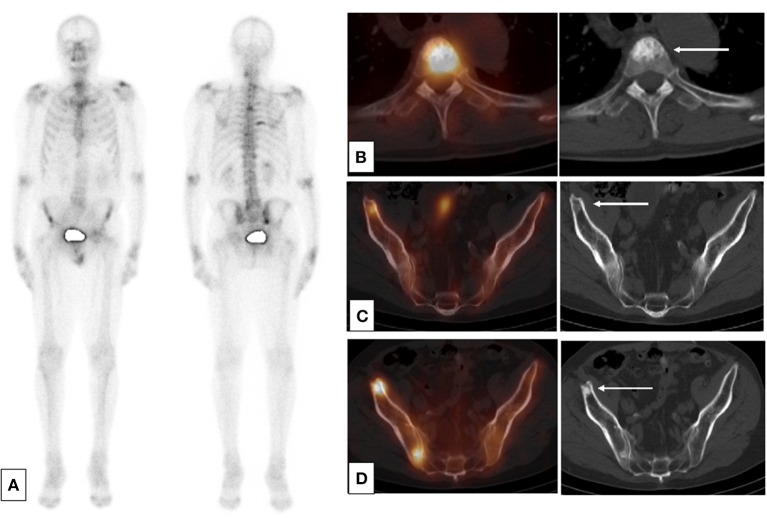
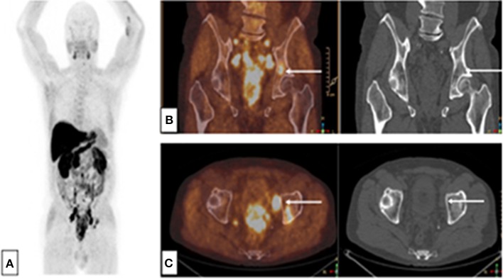
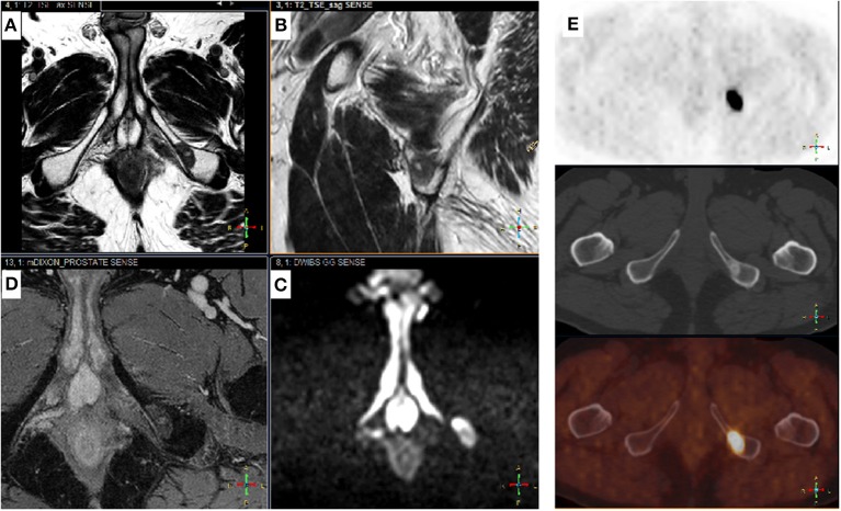
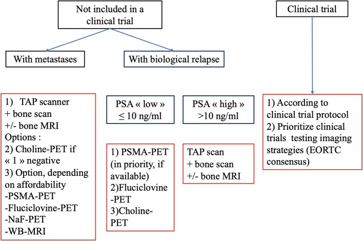
Background: Initial staging and assessment of treatment activity in metastatic prostate cancer (PCa) patients is controversial. Indications for the various available imaging modalities are not well-established due to rapid advancements in imaging and treatment. Methods: We conducted a critical literature review of the main imaging abnormalities that suggest a diagnosis of metastasis in localized and locally advanced PCa or in cases of biological relapse. We also assessed the role of the various imaging modalities available in routine clinical practice for the detection of metastases and response to treatment in metastatic PCa patients. Results: In published clinical trials, the most commonly used imaging modalities for the detection and evaluation of therapeutic response are bone scan, abdominopelvic computed tomography (CT), and pelvic and bone magnetic resonance imaging (MRI). For the detection and follow-up of metastases during treatment, modern imaging techniques i.e., choline-positron emission tomography (PET), fluciclovine-PET, or Prostate-specific membrane antigen (PSMA)-PET provide better sensitivity and specificity. This is particularly the case of fluciclovine-PET and PSMA-PET in cases of biochemical recurrence with low values of prostate specific antigen. Conclusions: In routine clinical practice, conventional imaging still have a role, and communication between imagers and clinicians should be encouraged. Present and future clinical trials should use modern imaging methods to clarify their usage.

Keywords: MRI; PSMA-PET; bone scan; choline-PET; fluciclovine-PET; prostate cancer; staging.


Figures

Figure 1
Figure 1
SPECT-CT: condensation of bone lesions under treatment. Occurrence of a prostate adenocarcinoma, Gleason 6 (3 + 3) on a biopsy, unoperated, treated by hormone therapy and HIFU therapy in a patient. Progressive re-elevation of the PSA 2 years after the cessation of hormone therapy. (A) Baseline planar bone scan (B,C) Baseline SPECT-CT and CT (axial slices) of lesions of T4 (B) and right ilium (C). (D) SPECT-CT and CT after 1 year of treatment by leuprorelin acetate showing an osteosclerotic reaction in the right ilium.
Figure 2
Figure 2
Choline-PET-CT: Single bone lesion of the left acetabulum, without CT abnormality. Initial assessment of a patient with immediately metastatic prostate adenocarcinoma with bone and node lesions. Gleason 8 (4 + 4), cT3, PSA = 36 ng/mL, (A) MIP reconstruction, (B) PET-CT and CT frontal slices, (C) PET-CT and CT axial slices.
Figure 3
Figure 3
Complementary Choline-TEP and pelvic MRI. Patient with a history of Gleason 7 (4 + 3) prostatic adenocarcinoma treated by radical prostatectomy 9 years ago (pT3aN0M0R1). PSA recurrence 5 years later followed by EBRT, external beam radiation therapy. Three years later, rising PSA from 0.5 to 1.4 ng/mL in 6 months. Prostate MRI performed before 18-F choline-PET-CT shows no sign of recurrence in the prostatectomy bed, but detected a 17-mm suspicious bone lesion in the left ischiopubic branch. Typical signal on axial (A) and sagittal (B) T2-w images were low and homogeneous; high on b-1000 axial diffusion-weighted images (DWI) (C) and high on late T1-w dynamic contrast-enhanced (DCE) images (D), corresponding to high cellular density and hypervascularization, respectively. 18F choline-PET-CT (E): axial slices of the pelvis showing an osteosclerotic lesion on the right ischiopubic branch with high uptake of the tracer. From top to bottom: PET, CT, PET-CT.
Figure 4
Figure 4
Flow chart of recommended imaging in evaluating metastatic PCa in routine practice.

References

    1. Bray F, Ferlay J, Soerjomataram I, Siegel RL, Torre LA, Jemal A. Global cancer statistics 2018: GLOBOCAN estimates of incidence and mortality worldwide for 36 cancers in 185 countries. CA Cancer J Clin. (2018) 68:394–424. 10.3322/caac.21492 - DOI - PubMed
    1. Sartor O, de Bono JS. Metastatic prostate cancer. N Engl J Med. (2018) 378:1653–4. 10.1056/NEJMra1701695 - DOI - PubMed
    1. Gillessen S, Omlin A, Attard G, de Bono JS, Efstathiou E, Fizazi K, et al. Management of patients with advanced prostate cancer: recommendations of the St Gallen Advanced Prostate Cancer Consensus Conference (APCCC) 2015. Ann Oncol. (2015). 26:1589–604. 10.1093/annonc/mdv360 - DOI - PMC - PubMed
    1. Gillessen S, Attard G, Beer TM, Beltran H, Bossi A, Bristow R, et al. Management of patients with advanced prostate cancer: the Report of the Advanced Prostate Cancer Consensus Conference APCCC 2017. Eur Urol. (2017). 73:178–211. 10.1016/j.eururo.2017.06.002 - DOI - PubMed
    1. Fanti S, Minozzi S, Antoch G, Banks I, Briganti A, Carrio I, et al. . Consensus on molecular imaging and theranostics in prostate cancer. Lancet Oncol. (2018) 19:e696–708. 10.1016/S1470-2045(18)30604-1 - DOI - PubMed

Publication types