

Structure, Properties, and Function of Glycosomes in Trypanosoma cruzi
- PMID: 32083023
- PMCID: PMC7005584
- DOI: 10.3389/fcimb.2020.00025
Structure, Properties, and Function of Glycosomes in Trypanosoma cruzi
Abstract
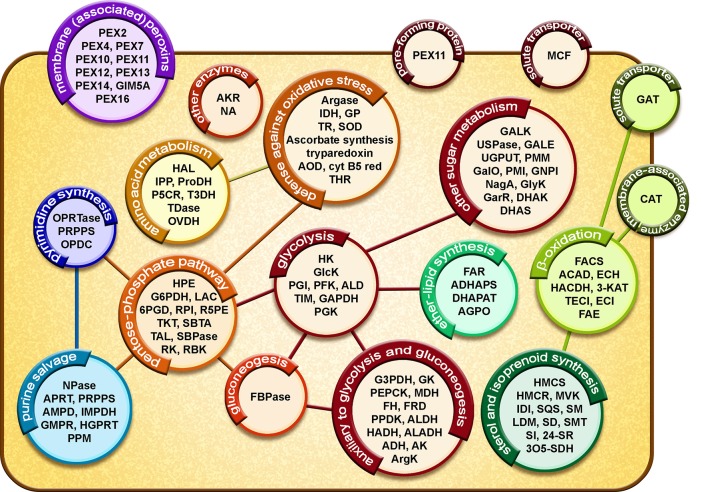
Glycosomes are peroxisome-related organelles that have been identified in kinetoplastids and diplonemids. The hallmark of glycosomes is their harboring of the majority of the glycolytic enzymes. Our biochemical studies and proteome analysis of Trypanosoma cruzi glycosomes have located, in addition to enzymes of the glycolytic pathway, enzymes of several other metabolic processes in the organelles. These analyses revealed many aspects in common with glycosomes from other trypanosomatids as well as features that seem specific for T. cruzi. Their enzyme content indicates that T. cruzi glycosomes are multifunctional organelles, involved in both several catabolic processes such as glycolysis and anabolic ones. Specifically discussed in this minireview are the cross-talk between glycosomal metabolism and metabolic processes occurring in other cell compartments, and the importance of metabolite translocation systems in the glycosomal membrane to enable the coordination between the spatially separated processes. Possible mechanisms for metabolite translocation across the membrane are suggested by proteins identified in the organelle's membrane-homologs of the ABC and MCF transporter families-and the presence of channels as inferred previously from the detection of channel-forming proteins in glycosomal membrane preparations from the related parasite T. brucei. Together, these data provide insight in the way in which different parts of T. cruzi metabolism, although uniquely distributed over different compartments, are integrated and regulated. Moreover, this information reveals opportunities for the development of drugs against Chagas disease caused by these parasites and for which currently no adequate treatment is available.
Keywords: biogenesis; drug discovery; glycolysis; glycosomes; metabolic networks; metabolite transport; peroxisomes; trypanosomes.
Copyright © 2020 Quiñones, Acosta, Gonçalves, Motta, Gualdrón-López and Michels.
Figures
Similar articles
-
Isolation of Glycosomes from Trypanosoma brucei.Methods Mol Biol. 2023;2643:33-45. doi: 10.1007/978-1-0716-3048-8_3. Methods Mol Biol. 2023. PMID: 36952176
-
The Trypanosome UDP-Glucose Pyrophosphorylase Is Imported by Piggybacking into Glycosomes, Where Unconventional Sugar Nucleotide Synthesis Takes Place.mBio. 2021 Jun 29;12(3):e0037521. doi: 10.1128/mBio.00375-21. Epub 2021 May 28. mBio. 2021. PMID: 34044588 Free PMC article.
-
Delineating transitions during the evolution of specialised peroxisomes: Glycosome formation in kinetoplastid and diplonemid protists.Front Cell Dev Biol. 2022 Sep 12;10:979269. doi: 10.3389/fcell.2022.979269. eCollection 2022. Front Cell Dev Biol. 2022. PMID: 36172271 Free PMC article.
-
Metabolic functions of glycosomes in trypanosomatids.Biochim Biophys Acta. 2006 Dec;1763(12):1463-77. doi: 10.1016/j.bbamcr.2006.08.019. Epub 2006 Aug 24. Biochim Biophys Acta. 2006. PMID: 17023066 Review.
-
Glycosomes: A comprehensive view of their metabolic roles in T. brucei.Int J Biochem Cell Biol. 2017 Apr;85:85-90. doi: 10.1016/j.biocel.2017.01.015. Epub 2017 Feb 6. Int J Biochem Cell Biol. 2017. PMID: 28179189 Review.
Cited by
-
3-Bromopyruvate Impairs Mitochondrial Function in Trypanosoma cruzi.Pathogens. 2025 Jun 25;14(7):631. doi: 10.3390/pathogens14070631. Pathogens. 2025. PMID: 40732679 Free PMC article.
-
Trypanosoma cruzi pathogenicity involves virulence factor expression and upregulation of bioenergetic and biosynthetic pathways.Virulence. 2022 Dec;13(1):1827-1848. doi: 10.1080/21505594.2022.2132776. Virulence. 2022. PMID: 36284085 Free PMC article.
-
Towards solving the mystery of peroxisomal matrix protein import.Trends Cell Biol. 2024 May;34(5):388-405. doi: 10.1016/j.tcb.2023.08.005. Epub 2023 Sep 22. Trends Cell Biol. 2024. PMID: 37743160 Free PMC article. Review.
-
Energy metabolism in Trypanosoma cruzi: the validated and putative bioenergetic and carbon sources.mBio. 2025 Jun 11;16(6):e0221524. doi: 10.1128/mbio.02215-24. Epub 2025 May 20. mBio. 2025. PMID: 40391931 Free PMC article. Review.
-
Translating the Arabidopsis thaliana Peroxisome Proteome Insights to Solanum lycopersicum: Consensus Versus Diversity.Front Cell Dev Biol. 2022 Jul 13;10:909604. doi: 10.3389/fcell.2022.909604. eCollection 2022. Front Cell Dev Biol. 2022. PMID: 35912119 Free PMC article. Review.
References
-
- Acosta H., Dubourdieu M., Quiñones W., Cáceres A., Bringaud F., Concepción J. L. (2004). Pyruvate phosphate dikinase and pyrophosphate metabolism in the glycosome of Trypanosoma cruzi epimastigotes. Comp. Biochem. Physiol. B Biochem. Mol. Biol. 138, 347–356. 10.1016/j.cbpc.2004.04.017 - DOI - PubMed
Publication types
MeSH terms
LinkOut - more resources
Full Text Sources
Medical