Review
. 2020 Jan 31:8:22.
doi: 10.3389/fcell.2020.00022. eCollection 2020.

The Generation of Dynein Networks by Multi-Layered Regulation and Their Implication in Cell Division

Affiliations
Review

The Generation of Dynein Networks by Multi-Layered Regulation and Their Implication in Cell Division

Takayuki Torisawa et al. Front Cell Dev Biol. .

Abstract

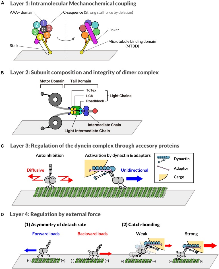
Cytoplasmic dynein-1 (hereafter referred to as dynein) is a major microtubule-based motor critical for cell division. Dynein is essential for the formation and positioning of the mitotic spindle as well as the transport of various cargos in the cell. A striking feature of dynein is that, despite having a wide variety of functions, the catalytic subunit is coded in a single gene. To perform various cellular activities, there seem to be different types of dynein that share a common catalytic subunit. In this review, we will refer to the different kinds of dynein as "dyneins." This review attempts to classify the mechanisms underlying the emergence of multiple dyneins into four layers. Inside a cell, multiple dyneins generated through the multi-layered regulations interact with each other to form a network of dyneins. These dynein networks may be responsible for the accurate regulation of cellular activities, including cell division. How these networks function inside a cell, with a focus on the early embryogenesis of Caenorhabditis elegans embryos, is discussed, as well as future directions for the integration of our understanding of molecular layering to understand the totality of dynein's function in living cells.

Keywords: C. elegans; centrosome positioning; cytoplasmic dynein-1; microtubule; motor activity.


Figures

FIGURE 1
FIGURE 1
Multi-layered regulatory mechanism of cytoplasmic dynein-1. (A) Layer 1: distinct regions of the C-terminal motor domain of dynein heavy chain subunit are shown with different colors. The number shows distinct AAA motifs. “C” indicates the C-sequence. The stalk and microtubule binding domain extending from AAA4 are shown in yellow. The linker connected to AAA1 is depicted as a purple bar. (B) Layer 2: distinct subunits of dynein complex are shown with different colors. Red represents the dimerization domain of the heavy chain. (C) Layer 3: Through the interaction with adaptor proteins, the localization and activity of dynein is regulated. A single complex of dynein that forms a phi-particle is auto-inhibited and diffuses along the microtubule (left). In contrast, dynein associated with dynactin and adaptor protein moves unidirectionally (right). (D) Layer 4: Dynein activity is also regulated by forces. When dynein is pulled toward the plus-end of microtubules, it binds more strongly to microtubules compared to dynein without force or pulled toward the minus-end (1). Additionally, the dynein dissociation rate decreases in the presence of high loads (2).
FIGURE 2
FIGURE 2
Intracellular localization and function of dynein during the first cell division of C. elegans one-cell embryo (A–I, in temporal order, see also text). Intracellular localization of the heavy chain subunit of dynein (DHC-1) is shown in dark and light gray (Gonczy et al., 1999). Nuclei and chromosomes are shown in blue. The movement of the centrosomes driven by dynein are denoted by red arrows. See Table 1 for the references describing the involvement of dynein in each process.

References

    1. Ally S., Larson A. G., Barlan K., Rice S. E., Gelfand V. I. (2009). Opposite-polarity motors activate one another to trigger cargo transport in live cells. J. Cell Biol. 187 1071–1082. 10.1083/jcb.200908075 - DOI - PMC - PubMed
    1. Barbar E., Nyarko A. (2015). Polybivalency and disordered proteins in ordering macromolecular assemblies. Semin. Cell Dev. Biol. 37 20–25. 10.1016/j.semcdb.2014.09.016 - DOI - PMC - PubMed
    1. Barbosa D. J., Duro J., Prevo B., Cheerambathur D. K., Carvalho A. X., Gassmann R. (2017). Dynactin binding to tyrosinated microtubules promotes centrosome centration in C. elegans by enhancing dynein-mediated organelle transport. PLoS Genet. 13:e1006941. 10.1371/journal.pgen.1006941 - DOI - PMC - PubMed
    1. Barkus R. V., Klyachko O., Horiuchi D., Dickson B. J., Saxton W. M. (2008). Identification of an axonal kinesin-3 motor for fast anterograde vesicle transport that facilitates retrograde transport of neuropeptides. Mol. Biol. Cell 19 274–283. 10.1091/mbc.e07-03-0261 - DOI - PMC - PubMed
    1. Belyy V., Schlager M. A., Foster H., Reimer A. E., Carter A. P., Yildiz A. (2016). The mammalian dynein-dynactin complex is a strong opponent to kinesin in a tug-of-war competition. Nat. Cell Biol. 18 1018–1024. 10.1038/ncb3393 - DOI - PMC - PubMed

LinkOut - more resources