. 2019 Dec 30:2019:1324804.
doi: 10.1155/2019/1324804. eCollection 2019.

Anti-PLA2R1 Antibodies Containing Sera Induce In Vitro Cytotoxicity Mediated by Complement Activation

Affiliations

Anti-PLA2R1 Antibodies Containing Sera Induce In Vitro Cytotoxicity Mediated by Complement Activation

Maël Lateb et al. J Immunol Res. .

Abstract

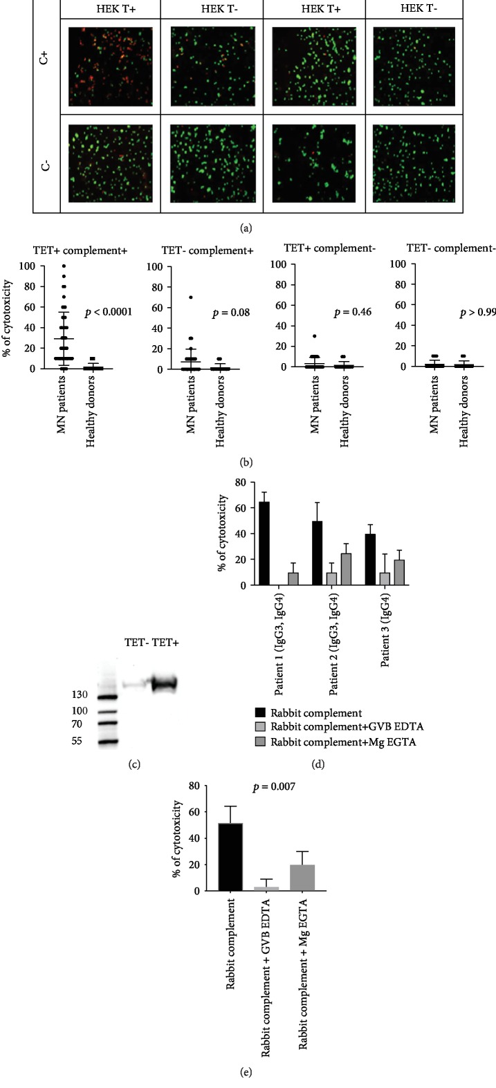
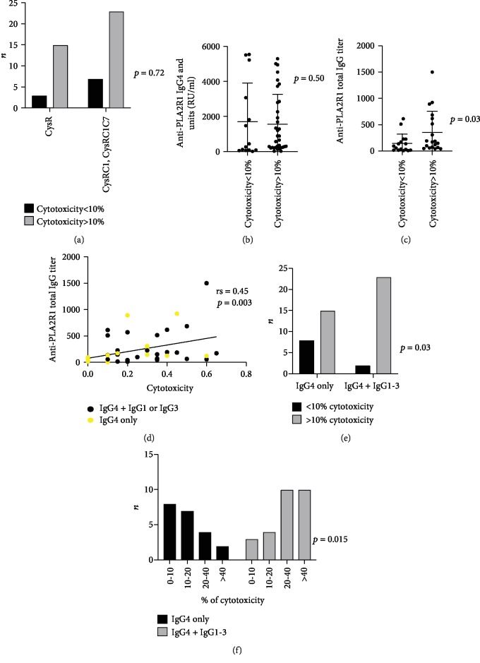
The phospholipase A2 receptor (PLA2R1) is the major autoantigen in idiopathic membranous nephropathy (MN). However, the pathogenic role of anti-PLA2R1 autoantibodies is unclear. Our aim was to evaluate the in vitro cytotoxicity of anti-PLA2R1 antibodies mediated by complement. Forty-eight patients with PLA2R1-related MN from the prospective cohort SOURIS were included. Anti-PLA2R1 titer, epitope profile, and anti-PLA2R1 IgG subclasses were characterized by ELISA. Cell cytotoxicity was evaluated by immunofluorescence in HEK293 cells overexpressing PLA2R1 incubated with patient or healthy donor sera in the presence or absence of rabbit complement or complement inhibitors. Mean cytotoxicity of anti-PLA2R1 sera for HEK293 cells overexpressing PLA2R1 was 2 ± 2%, which increased to 24 ± 6% after addition of rabbit complement (p < 0.001) (n = 48). GVB-EDTA, which inhibits all complement activation pathways, completely blocked cell cytotoxicity, whereas Mg-EGTA, which only inhibits the classical and lectin pathways, highly decreased suggesting a limited role of the alternative pathway. A higher diversity of IgG subclasses beyond IgG4 and high titer of total IgG anti-PLA2R1 were associated with increased cytotoxicity (p = 0.01 and p = 0.03 respectively). In a cohort of 37 patients treated with rituximab, high level of complement-mediated cytotoxicity was associated with less and delayed remission at month 6 after rituximab therapy (5/12 vs. 20/25 (p = 0.03) in 8.5 months ± 4.4 vs. 4.8 ± 4.0 (p = 0.02)). Kaplan-Meier analysis demonstrated that high level of cytotoxicity (≥40%) (p = 0.005), epitope spreading (defined by immunization beyond the immunodominant CysR domain) (p = 0.002), and high titer of anti-PLA2R1 total IgG (p = 0.01) were factors of poor renal prognosis. Anti-PLA2R1 antibodies containing sera can induce in vitro cytotoxicity mediated by complement activation, and the level of cytotoxicity increases with the diversity and the titer of anti-PLA2R1 IgG subclasses. These patients with high level of complement-mediated cytotoxicity could benefit from adjuvant therapy using complement inhibitor associated with rituximab to induce earlier remission and less podocyte injury.


Conflict of interest statement

The authors declare that they have no conflicts of interest.

Figures

Figure 1
Figure 1
Description of samples with anti-PLA2R1 antibodies tested. (a) Epitope spreading profiles of patients at baseline (n = 48) and at last observation (n = 36). Follow-up was missing for 12 patients. C1: CTLD1; C7: CTLD7. CysR: immunized against CysR domain alone; CysRC1: immunized against CysR and CTLD1 domains; CysRC1C7: immunized against CysR, CTLD1, and CTLD7 domains. (b) IgG subclass profiles of patients at baseline (n = 48). (c) IgG subclass profiles of patients according to disease course: first course or relapse (n = 48). Note that patients with the first course of MN have a larger diversity of IgG subclasses (p = 0.02).
Figure 2
Figure 2
Anti-PLA2R1-mediated cytotoxicity depends on complement in immunofluorescence cytotoxicity assay. (a) Anti-PLA2R1-mediated cytotoxicity depending on complement for a PLA2R1-positive serum and a healthy donor serum. HEK T+: HEK293T-REx cells transfected with PLA2R1 and induced with tetracycline to overexpress PLA2R1. HEK T-: HEK293T-REx cells transfected with PLA2R1 but not induced with tetracycline. C+: addition of rabbit complement. C-: no addition of rabbit complement. Dead cells appear in red; living cells appear in green. (b) Anti-PLA2R1 complement-mediated cytotoxicity in a cohort of 48 patients and 20 healthy donors in different conditions. Note a low level of cytotoxicity in noninduced HEK that tended to be significant (p = 0.08) caused by a minimal expression of PLA2R1 even in noninduced cells, as determined by western blot (c). All samples were tested using the same batch of HEK293 cells. A minimum of 50 cells per well was necessary for reading. (c) Expression of PLA2R1 in noninduced and induced HEK293 cells with tetracycline. Note a minimal expression of PLA2R1 in noninduced cells. (d) Analysis of complement activation pathways involved in anti-PLA2R1-mediated cytotoxicity for 3 PLA2R1-positive patients using two inhibitors of complement pathways. Patients 1 and 2 were positive for both IgG3 and IgG4 anti-PLA2R1 antibodies, while patient 3 was only positive for IgG4 anti-PLA2R1. An excess of GVB-EDTA (inhibitor of the three pathways of the complement) or Mg-EGTA (inhibitor of the classical and lectin pathways) was added in serum + complement and the complement-mediated cytotoxicity was measured. All samples were tested using the same batch of HEK293 cells. A minimum of 50 cells per well was necessary for reading. (e) Analysis of complement activation pathways involved in anti-PLA2R1-mediated cytotoxicity for 3 PLA2R1-positive patients. Note that GVB-EDTA strongly inhibits anti-PLA2R1-mediated cytotoxicity in all 3 patients, while Mg-EGTA, which is known to inhibit only the classical and lectin pathways, inhibits only partially the anti-PLA2R1-mediated cytotoxicity, suggesting a potential activation of the alternative pathway in some serum samples. A minimum of 50 cells per well was necessary for reading. HEK293 T-REx cells transfected with PLA2R1 and induced with tetracycline to express PLA2R1 were used. Negative controls (noninduced HEK T-REx cells) are not shown. All cytotoxicity assays were performed using the same batch of HEK293 T-Rex cells. A minimum of 50 cells per well was necessary for reading.
Figure 3
Figure 3
Predictive factors of anti-PLA2R1-mediated cytotoxicity in immunofluorescence cytotoxicity assay. (a) Relationship between anti-PLA2R1-induced cytotoxicity mediated by complement and epitope spreading profile at baseline (n = 48). CysR: immunized against CysR domain alone; CysRC1: immunized against CysR and CTLD1 domains; CysRC1C7: immunized against CysR, CTLD1, and CTLD7domains. (b) Relationship between anti-PLA2R1-induced cytotoxicity mediated by complement and Anti-PLA2R1 IgG4 (RU/ml) titers at baseline (n = 48). (c) Relationship between anti-PLA2R1-induced cytotoxicity mediated by complement and Anti-PLA2R1 total IgG titer at baseline (n = 48). (d) Correlation between Anti-PLA2R1 total IgG titer and complement-mediated cytotoxicity (n = 48). (e) Anti-PLA2R1-induced cytotoxicity mediated by complement according to anti-PLA2R1 IgG subclasses at baseline (n = 48). (f) Anti-PLA2R1-induced cytotoxicity mediated by complement regrouped by levels of cytotoxicity according to anti-PLA2R1 IgG subclasses at baseline (n = 48). Note that patients with high level of cytotoxicity (≥40%) are predominantly IgG4+IgG1-IgG3.
Figure 4
Figure 4
Factors associated with remission. Renal event is defined by remission (partial or complete) one year after diagnosis of MN. (a) Percent of patients achieving remission according to the level of complement-mediated cytotoxicity (n = 37). (b) Percent of patients achieving remission according to epitope spreading profile (n = 37). (c) Percent of patients achieving remission according to the level of total IgG anti-PLA2R1 (n = 37).

References

    1. Simon P., Ramee M. P., Boulahrouz R., et al. Epidemiologic data of primary glomerular diseases in western France. Kidney International. 2004;66(3):905–908. doi: 10.1111/j.1523-1755.2004.00834.x. - DOI - PubMed
    1. Ronco P., Debiec H. Pathophysiological advances in membranous nephropathy: time for a shift in patient’s care. The Lancet. 2015;385(9981):1983–1992. doi: 10.1016/S0140-6736(15)60731-0. - DOI - PubMed
    1. Beck L. H., Jr., Bonegio R. G. B., Lambeau G., et al. M-type phospholipase A2 receptor as target antigen in idiopathic membranous nephropathy. The New England Journal of Medicine. 2009;361(1):11–21. doi: 10.1056/NEJMoa0810457. - DOI - PMC - PubMed
    1. Tomas N. M., Beck L. H., Jr., Meyer-Schwesinger C., et al. Thrombospondin type-1 domain-containing 7A in idiopathic membranous nephropathy. The New England Journal of Medicine. 2014;371(24):2277–2287. doi: 10.1056/NEJMoa1409354. - DOI - PMC - PubMed
    1. Zaghrini C., Seitz-Polski B., Justino J., et al. Novel ELISA for thrombospondin type 1 domain-containing 7A autoantibodies in membranous nephropathy. Kidney International. 2019;95(3):666–679. doi: 10.1016/j.kint.2018.10.024. - DOI - PubMed

MeSH terms

LinkOut - more resources