

Progressive brainstem pathology in motor neuron diseases: Imaging data from amyotrophic lateral sclerosis and primary lateral sclerosis
- PMID: 32083157
- PMCID: PMC7016370
- DOI: 10.1016/j.dib.2020.105229
Progressive brainstem pathology in motor neuron diseases: Imaging data from amyotrophic lateral sclerosis and primary lateral sclerosis
Abstract
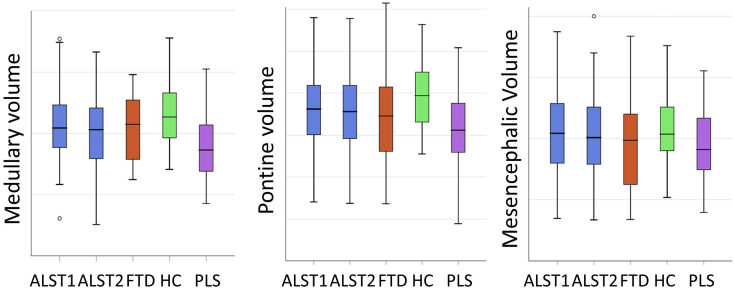
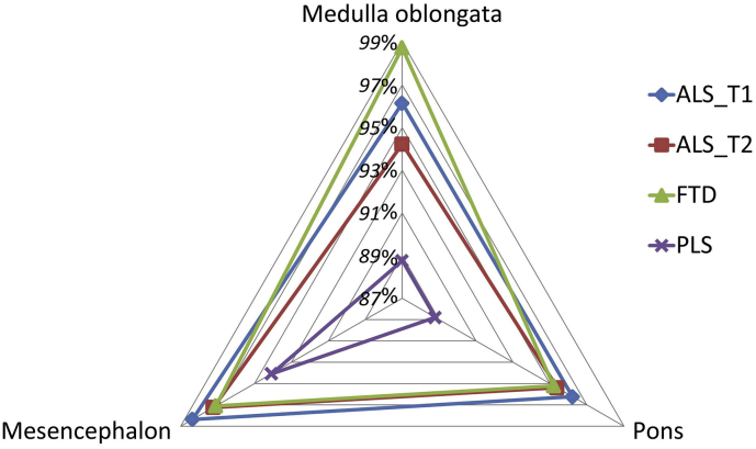
A standardised, single-centre, longitudinal imaging protocol was used to evaluate longitudinal brainstem alterations in 100 patients with amyotrophic lateral sclerosis (ALS) with reference to 33 patients with primary lateral sclerosis (PLS), 30 patients with frontotemporal dementia (FTD) and 100 healthy controls. "Brainstem pathology in amyotrophic lateral sclerosis and primary lateral sclerosis: A longitudinal neuroimaging study" [1] ALS patients were scanned twice; 4 months apart. T1-weighted imaging data were acquired on a 3 T Philips Achieva MRI system, using a 3D Inversion Recovery prepared Spoiled Gradient Recalled echo (IR-SPGR) sequence. Raw MRI data underwent meticulous quality control before pre-processing. A Bayesian segmentation algorithm was utilised to parcellate the brainstem into the medulla oblongata, pons and mesencephalon before estimating the volume of each segment. Vertex-based shape analyses were carried out to characterise anatomical patterns of atrophy. Brainstem volume loss in ALS was dominated by medulla oblongata atrophy, but significant pontine pathology was also detected. Brainstem volume reductions were more significant in PLS than in ALS after correcting for demographic variables and total intracranial volume. Shape analyses revealed bilateral 'flattening' of the medullary pyramids in ALS compared to healthy controls. Our data demonstrate that computational neuroimaging readily detects brainstem pathology in vivo in both amyotrophic lateral sclerosis and primary lateral sclerosis.
Keywords: Amyotrophic lateral sclerosis; Brainstem; Frontotemporal dementia; Magnetic resonance imaging; Medulla oblongata; Mesencephalon; Pons; Primary lateral sclerosis.
© 2020 The Authors.
Figures

References
-
- Bede P., Chipika R.H., Finegan E., Li Hi Shing S., Doherty M.A., Hengeveld J.C., Vajda A., Hutchinson S., Donaghy C., McLaughlin R.L., Hardiman O. Brainstem pathology in amyotrophic lateral sclerosis and primary lateral sclerosis: a longitudinal neuroimaging study. NeuroImage Clinical. 2019;24:102054. Epub 2019/11/12. - PMC - PubMed
-
- Bede P., Finegan E. Revisiting the pathoanatomy of pseudobulbar affect: mechanisms beyond corticobulbar dysfunction. Amyotroph Lateral Scler. frontotemporal degeneration. 2018;19:4–6. Epub 2017/11/03. - PubMed
-
- Hardiman O., Doherty C.P., Elamin M., Bede P. Springer Cham Heidelberg New York Dordrecht London© Springer International Publishing Switzerland 2016. Springer International Publishing; 2016. Neurodegenerative disorders: a clinical guide. 2016; pp. 1–336.
LinkOut - more resources
Full Text Sources
Miscellaneous
