. 2020 Mar 27;40(3):BSR20192723.
doi: 10.1042/BSR20192723.

Down-regulation of microRNA-203a suppresses IL-1β-induced inflammation and cartilage degradation in human chondrocytes through Smad3 signaling

Affiliations

Down-regulation of microRNA-203a suppresses IL-1β-induced inflammation and cartilage degradation in human chondrocytes through Smad3 signaling

Yongbo An et al. Biosci Rep. .

Abstract

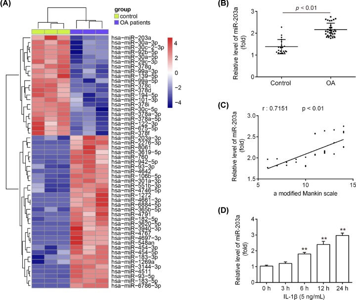
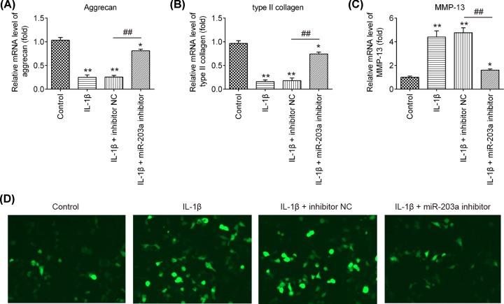
Osteoarthritis (OA) is a chronic and prevalent degenerative musculoskeletal disorder, which is characterized by articular cartilage degradation and joint inflammation. MicroRNA-203a (miR-203a) has been shown to be involved in multiple pathological processes during OA, but little is known about its function in chondrocyte extracellular matrix (ECM) degradation. In the present study, we aimed to elucidate the effects of miR-203a on articular cartilage degradation and joint inflammation. We observed that the miR-203a level was significantly up-regulated in OA tissues and in an in vitro model of OA, respectively. Inhibition of miR-203a significantly alleviated the interleukin (IL)-1β-induced inflammatory response and ECM degradation in chondrocytes. Moreover, mothers against decapentaplegic homolog 3 (Smad3), a key factor in maintaining chondrocyte homeostasis, was identified as a putative target of miR-203a in chondrocytes. More importantly, inhibition of Smad3 impaired the inhibitory effects of the miR-203a on IL-1β-induced inflammatory response and ECM degradation. Collectively, these results demonstrated that miR-203a may contribute to articular cartilage degradation of OA by targeting Smad3, suggesting a novel therapeutic target for the treatment of OA.

Keywords: ECM; Osteoarthritis; articular cartilage; inflammatory cytokines; miR-203a.


Conflict of interest statement

The authors declare that there are no competing interests associated with the manuscript.

Figures

Figure 1
Figure 1. The miR-203a level in human OA articular cartilage tissues and IL-1β-induced chondrocytes
(A) The microarray analysis was used to identify the miRNA expression profiles in human normal and OA articular cartilage tissues. Red or green color separately shows high or low expression in the heatmap. (B) The miR-203a level was determined by qRT-PCR in human normal (n=20) and OA articular cartilage tissues (n=30). (C) The miR-203a expression level was positively correlated with Mankin scale (r = 0.7151, P<0.01). (D) Chondrocytes were treated with IL-1β at 5 ng/ml for 0, 3, 6, 12, or 24 h, and the miR-203a level were detected using qRT-PCR. Data are presented as means ± SD of three individual experiments (**P<0.01 vs. control).
Figure 2
Figure 2. The effect of miR-203a down-regulation on the IL-1β-induced cell viability, apoptosis and inflammatory cytokines production
Chondrocytes transfected with miR-203a inhibitor and inhibitor NC and then treated with IL-1β at 5 ng/ml for 24 h. (A) The miR-203a mRNA level was measured by the RT-qPCR assay. (B) The CCK-8 assay was used to measure the cell viability. (C,D) Cell apoptosis was evaluated using flow cytometry. (EG) Inflammatory cytokines TNF-α, IL-1β and IL-6 in supernatant samples were detected using ELISA kits, respectively. Data are presented as means ± SD of three individual experiments (*P<0.05, **P<0.01 vs. control group, ##P<0.01 vs. IL-1β + inhibitor NC).
Figure 3
Figure 3. The effect of miR-203a down-regulation on the IL-1β-induced aggrecan, type II collagen and MMP-13
The chondrocytes were transfected with miR-203a inhibitor or inhibitor NC, and then exposed chondrocytes to IL-1β for 24 h. (AC) The mRNA levels of aggrecan, type II collagen and MMP-13 were analyzed using qRT-PCR. (D) The MMP-13 protein level was detected using immunofluorescence assay. Data are presented as means ± SD of three individual experiments (*P<0.05, **P<0.01 vs. control group, ##P<0.01 vs. IL-1β + inhibitor NC).
Figure 4
Figure 4. Smad3 is a direct target of miR-203a in chondrocytes
(A) The Smad3 3ʹ-UTR region containing the wild-type (wt) or mutant (mut) binding site for miR-203a. (B) The chondrocytes were co-transfected with the reporter construct (pMIR-Smad3-3ʹ-UTR or pMIR-Smad3-3ʹ-UTR) and miR-203a mimic/inhibitor or corresponding NC and the relative luciferase activity were measured (**P<0.01 vs mimic NC; ##P<0.01 vs. inhibitor NC). (C) The Smad3 mRNA level was detected after miR-203a mimics transfection by qRT-PCR. (D) The protein level of Smad3 was detected using Western blot analysis (**P<0.01 vs. mimic NC; ##P<0.01 vs inhibitor NC). β-actin was used as an internal control. (E) The Smad3 mRNA level was detected using qRT-PCR in the OA articular cartilage tissues (n=30) and normal tissues (n=20) (**P<0.01 vs. control). (F) The negative correlation between Smad3 and miR-203a levels in the OA articular cartilage tissues (r = −0.6998, P<0.01). Data are presented as means ± SD of three individual experiments.
Figure 5
Figure 5. Smad3 silencing rescues the effects of miR-203a down-regulation on IL-1β-stimulated chondrocytes
The chondrocytes were transfected with miR-203a inhibitor or were co-transfected with miR-203a inhibitor and si-Smad3 or si-Scramble, and then treated with IL-1β at 5 ng/ml for 24 h. (A) The Smad3 protein level was determined using Western blot analysis. (B) Cell viability was measured using CCK-8 assay. (C,D) The flow cytometry was conducted to measure cell apoptosis. (EG) Inflammatory cytokines TNF-α, IL-1β and IL-6 were detected using ELISA kits, respectively. (HJ) The mRNA levels of aggrecan, type II collagen and MMP-13 were analyzed using qRT-PCR. Data are presented as means ± SD of three individual experiments (*P<0.05, **P<0.01 vs. IL-1β group, ##P<0.01 vs. miR-203a inhibitor + si-Scramble).

References

    1. Taruc-Uy R.L. and Lynch S.A. (2013) Diagnosis and treatment of osteoarthritis. Prim. Care 40, 821–836 10.1016/j.pop.2013.08.003 - DOI - PubMed
    1. Qin J. et al. . (2012) TNF/TNFR signal transduction pathway-mediated anti-apoptosis and anti-inflammatory effects of sodium ferulate on IL-1β-induced rat osteoarthritis chondrocytes in vitro. Arthritis Res. Ther. 14, R242. - PMC - PubMed
    1. Sandell L.J. and Aigner T. (2001) Articular cartilage and changes in arthritis: cell biology of osteoarthritis. Arthritis Res. Ther. 3, 107 - PMC - PubMed
    1. Bartel D.P. (2009) MicroRNAs: target recognition and regulatory functions. Cell 136, 215–233 10.1016/j.cell.2009.01.002 - DOI - PMC - PubMed
    1. Swingler T.E. et al. . (2011) The expression and function of microRNAs in chondrogenesis and osteoarthritis. Arthritis Rheum. 64, 1909–1919 - PubMed

Publication types