Review
. 2020 Aug;61(7):664-679.
doi: 10.1002/em.22365. Epub 2020 Feb 29.

Methodologies for detecting environmentally induced DNA damage and repair

Affiliations
Review

Methodologies for detecting environmentally induced DNA damage and repair

Wentao Li et al. Environ Mol Mutagen. 2020 Aug.

Abstract

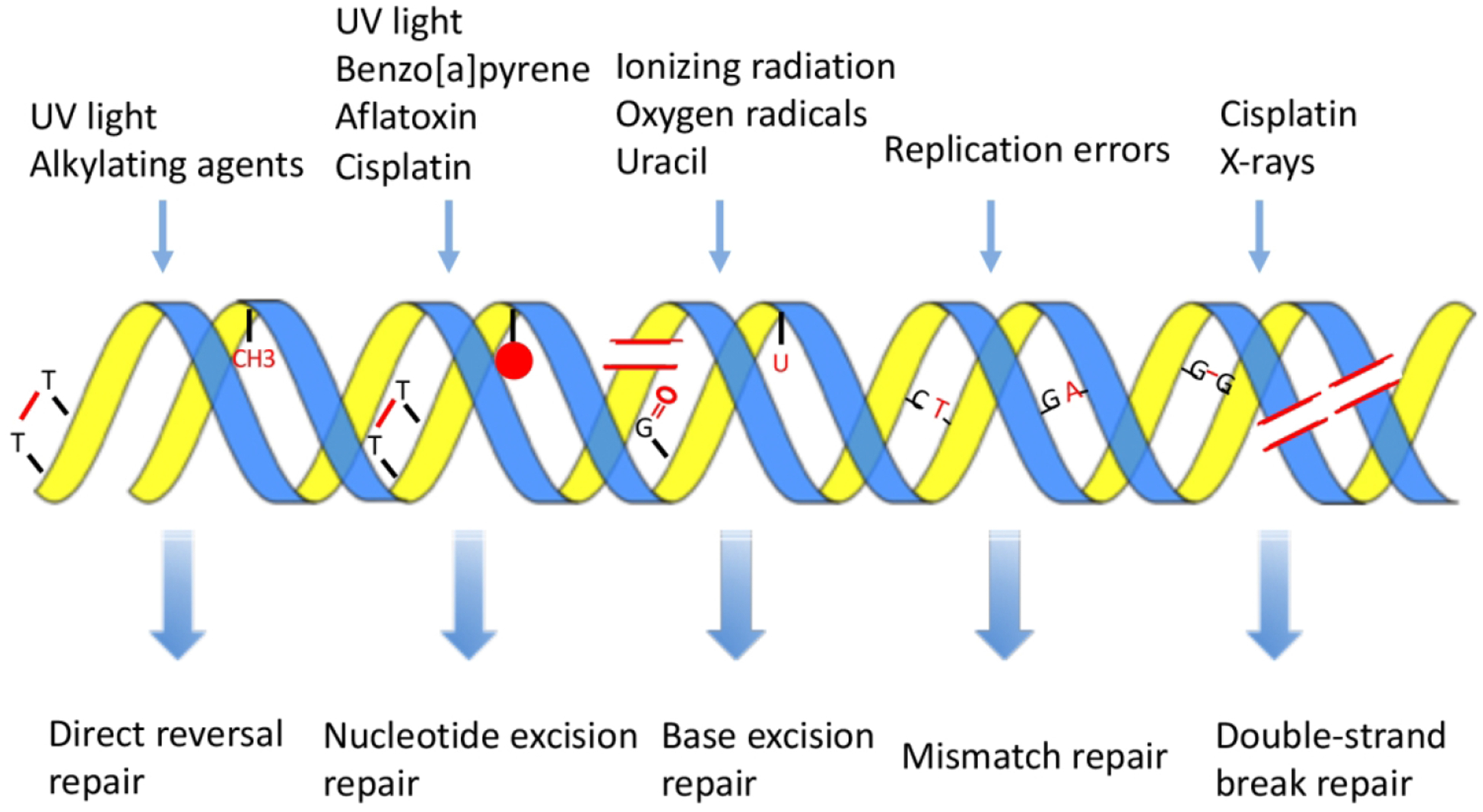
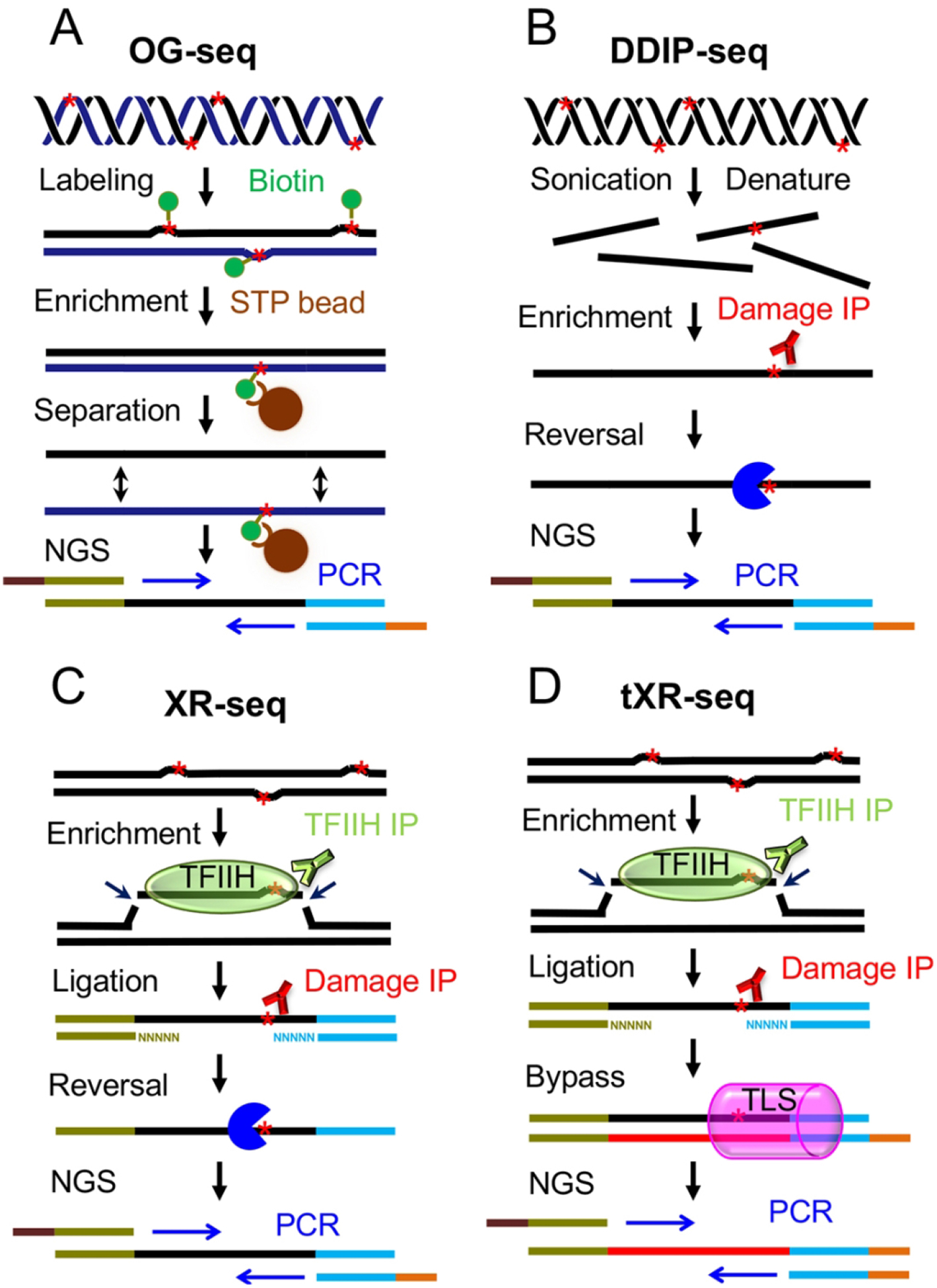
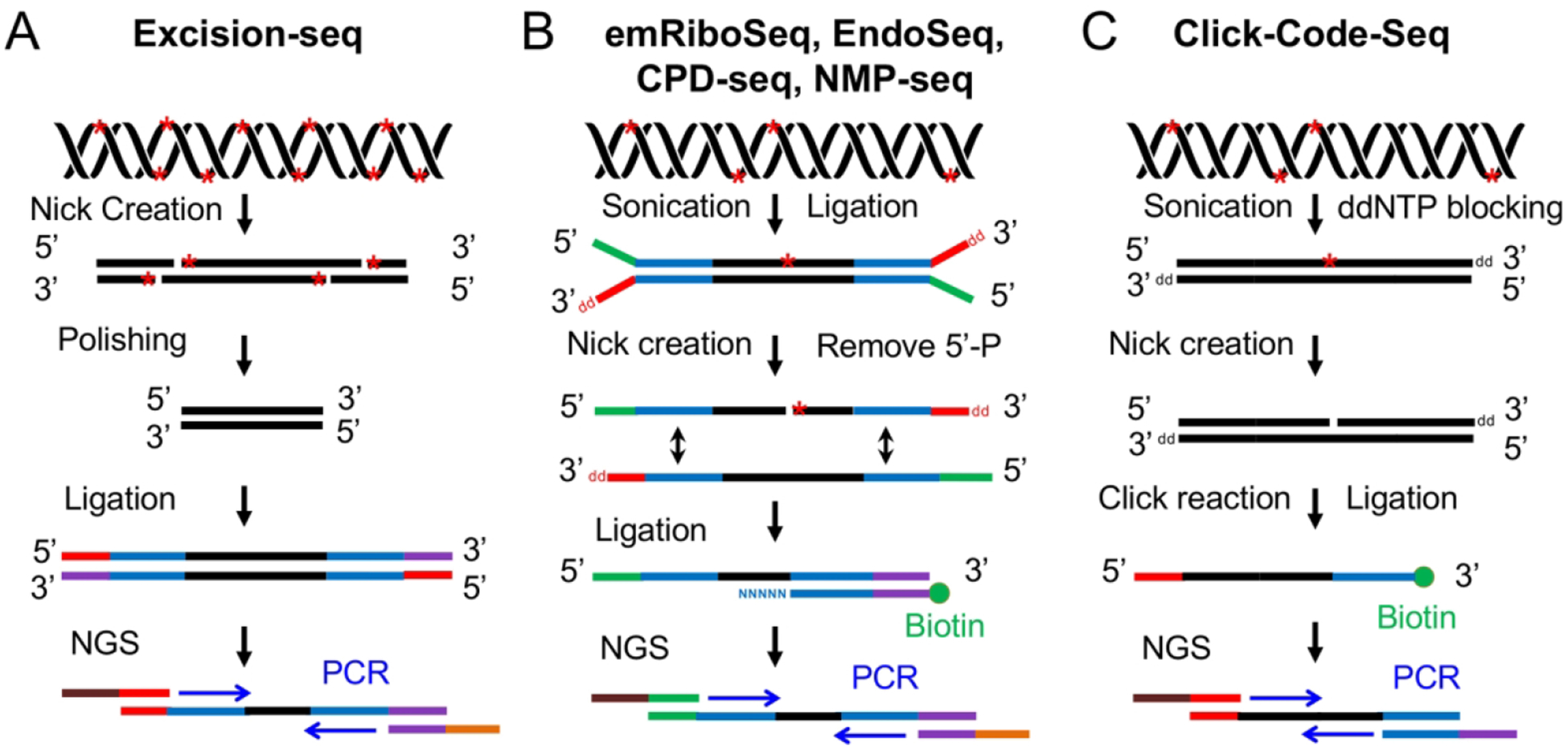
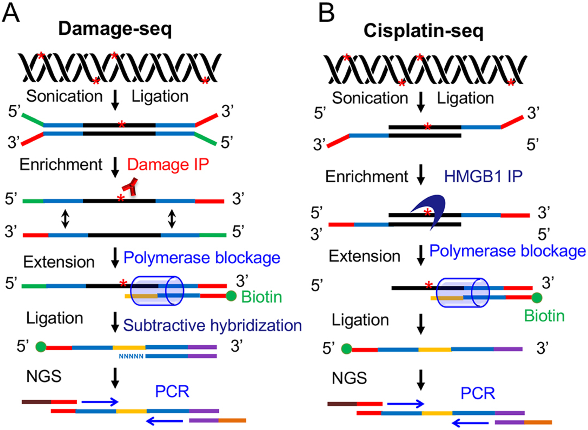
Environmental DNA damaging agents continuously challenge the integrity of the genome by introducing a variety of DNA lesions. The DNA damage caused by environmental factors will lead to mutagenesis and subsequent carcinogenesis if they are not removed efficiently by repair pathways. Methods for detection of DNA damage and repair can be applied to identify, visualize, and quantify the DNA damage formation and repair events, and they enable us to illustrate the molecular mechanisms of DNA damage formation, DNA repair pathways, mutagenesis, and carcinogenesis. Ever since the discovery of the double helical structure of DNA in 1953, a great number of methods have been developed to detect various types of DNA damage and repair. Rapid advances in sequencing technologies have facilitated the emergence of a variety of novel methods for detecting environmentally induced DNA damage and repair at the genome-wide scale during the last decade. In this review, we provide a historical overview of the development of various damage detection methods. We also highlight the current methodologies to detect DNA damage and repair, especially some next generation sequencing-based methods.

Keywords: DNA damage; DNA repair; mutagenesis; next-generation sequencing; third-generation sequencing.


Figures

Fig. 1.
Fig. 1.
Schematic of various DNA damaging agents-induced DNA damage and the corresponding repair pathways. The top panel shows the different types of DNA damaging agents. The various DNA lesions caused by DNA damaging agents are shown in red in the middle panel. The lower panel shows the corresponding repair pathways that are responsible for the removal of those DNA lesions listed in the middle panel. This figure is redrawn based on reference (Hoeijmakers 2001).
Fig. 2.
Fig. 2.
Schematic representation of four methods using the DNA damage enrichment and damage reversal or bypass-based strategy for detection of DNA damage and repair. (A) OG-seq for detection of 8-Oxoguanine (Ding et al. 2017). After fragmentation of genomic DNA containing oxidative damage, 8-Oxoguanines indicated by red stars are chemically labeled with biotin and enriched by streptavidin (STP) beads. Then, the double strands of DNA fragments are separated, and the strand (black) complementary to the damage containing strand (dark blue) will be amplified by PCR and sequenced by NGS. (B) Overview of DDIP-seq method (Alhegaili et al. 2019). The genomic DNA carrying CPDs represented by red stars is sonicated and denatured, and DNA strands containing damage are enriched by CPD immunoprecipitation. The CPDs are removed by repair enzymes (the dark blue pie), and then the strands are subjected to standard NGS library preparation procedure for sequencing. (C) XR-seq method for mapping repair of UV damage (Hu et al. 2015). The excised oligonucleotides carrying the UV damage indicated by red star are released in complex with TFIIH and enriched by TFIIH immunoprecipitation. After adapter ligation, the excised oligonucleotides are further purified by UV damage immunoprecipitation. The UV damage in the excised oligonucleotides are reversed by photolyases, and the excised oligonucleotides are amplified by PCR followed by NGS. Besides UV damage, the XR-seq can also be applied to detect repair of cisplatin-adducts which are reversed by sodium cyanide (Hu et al. 2016). (D) tXR-seq method for mapping repair of CPD and BPDE-DNA adducts (Li et al. 2017). The steps of DNA damage enrichment and ligation are similar to XR-seq. Instead of using damage reversal strategy, tXR-seq makes use of translesion DNA synthesis (TLS) polymerase (indicated by pink can) to bypass the DNA damage during one cycle of primer extension. The primer extension products are then amplified and subjected to NGS. This method can be applied to essentially all types of DNA damage that are removed by nucleotide excision repair.
Fig. 3.
Fig. 3.
Schematic of six NGS-based methods using nick creation and ligation-based strategy for detection of DNA damage. (A) Excision-seq method for detection of uracil and UV damage indicated by red stars in DNA (Bryan et al. 2014). The UDG and Endo IV are used to digest uracil, and UVDE and photolyases are applied to nick at sites of UV damage. After polishing, adapter ligation and PCR amplification, the library is subjected to NGS. (B) Overview of experimental workflow for emRiboSeq, EndoSeq, CPD-seq, NMP-seq (Ding et al. 2015; Mao et al. 2016; Mao et al. 2017). After sonication and ligation, the DNA damage (red stars) are recognized and digested by specific repair enzymes to create a nick 5’ to the lesion. Then, the 5’-P group is removed and the second adapter is ligated to the DNA fragments containing 3’-OH group. Following ligation, the ligation products are amplified by PCR and subjected to NGS. (C) Click-Code-Seq method for detection of oxidative damage (Wu et al. 2018). After sonication and ddNTP blocking, one nucleotide gap is created by FPG and APE1 repair enzymes. Then, a prop-dGTP is incorporated to fill the gap and a 5’-azido-modified code sequence is ligated to the 3’ end by click reaction followed by the 5’ end adaptor ligation. The ligation products are amplified and sequenced by NGS.
Fig. 4.
Fig. 4.
Schematic of two methods using DNA damage enrichment and primer extension-based strategy for detection of DNA damage. (A) Damage-seq method for detection of any type of damage that can block the DNA polymerase (Hu et al. 2016; Hu et al. 2017a). After sonication, ligation and denaturation, the DNA strands carrying damage are enriched by damage immunoprecipitation followed by one cycle of primer extension in which the high-fidelity DNA polymerase stops before the lesion. Following a subtractive hybridization step, the products of primer extension are then amplified by PCR for subsequent NGS. (B) Cisplatin-seq method for detection of cisplatin-DNA adducts (Shu et al. 2016). Following sonication and ligation, the DNA fragments containing cisplatin-DNA adducts are enriched by HMGB1 domain A immunoprecipitation followed by primer extension step which is similar to Damage-seq. Then, the primer extension products are ligated to a second adapter and amplified by PCR for the following NGS.

References

    1. Aaronson SA, Lytle CD. 1970. Decreased host cell reactivation of irradiated SV40 virus in xeroderma pigmentosum. Nature 228(5269):359–361. - PubMed
    1. Adar S, Hu J, Lieb JD, Sancar A. 2016. Genome-wide kinetics of DNA excision repair in relation to chromatin state and mutagenesis. Proc Natl Acad Sci U S A 113(15):E2124–2133. - PMC - PubMed
    1. Adebali O, Chiou YY, Hu J, Sancar A, Selby CP. 2017a. Genome-wide transcription-coupled repair in Escherichia coli is mediated by the Mfd translocase. Proc Natl Acad Sci U S A 114(11):E2116–E2125. - PMC - PubMed
    1. Adebali O, Sancar A, Selby CP. 2017b. Mfd translocase is necessary and sufficient for transcription-coupled repair in Escherichia coli. J Biol Chem 292(45):18386–18391. - PMC - PubMed
    1. Alhegaili AS, Ji Y, Sylvius N, Blades MJ, Karbaschi M, Tempest HG, Jones GDD, Cooke MS. 2019. Genome-Wide Adductomics Analysis Reveals Heterogeneity in the Induction and Loss of Cyclobutane Thymine Dimers across Both the Nuclear and Mitochondrial Genomes. Int J Mol Sci 20(20). - PMC - PubMed

Publication types