. 2020 Apr 6;48(6):3379-3394.
doi: 10.1093/nar/gkaa104.

A memory switch for plant synthetic biology based on the phage ϕC31 integration system

Affiliations

A memory switch for plant synthetic biology based on the phage ϕC31 integration system

Joan Miquel Bernabé-Orts et al. Nucleic Acids Res. .

Abstract

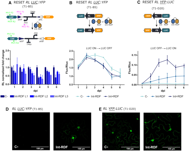
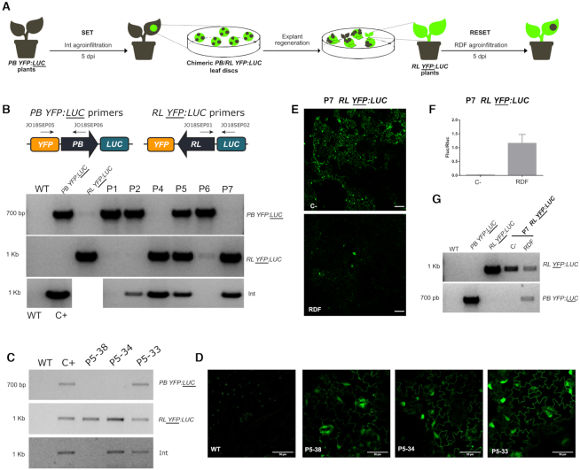
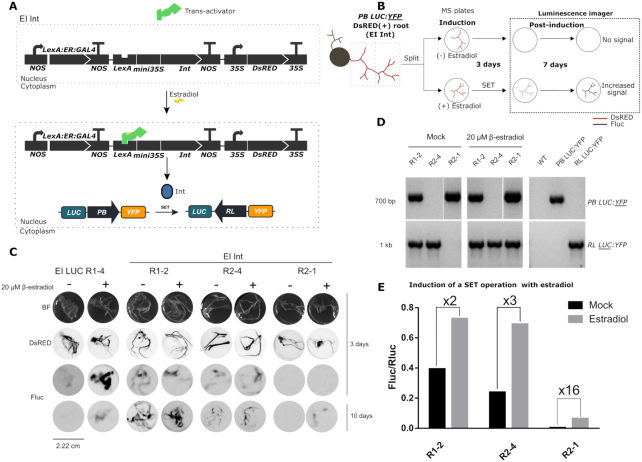
Synthetic biology has advanced from the setup of basic genetic devices to the design of increasingly complex gene circuits to provide organisms with new functions. While many bacterial, fungal and mammalian unicellular chassis have been extensively engineered, this progress has been delayed in plants due to the lack of reliable DNA parts and devices that enable precise control over these new synthetic functions. In particular, memory switches based on DNA site-specific recombination have been the tool of choice to build long-term and stable synthetic memory in other organisms, because they enable a shift between two alternative states registering the information at the DNA level. Here we report a memory switch for whole plants based on the bacteriophage ϕC31 site-specific integrase. The switch was built as a modular device made of standard DNA parts, designed to control the transcriptional state (on or off) of two genes of interest by alternative inversion of a central DNA regulatory element. The state of the switch can be externally operated by action of the ϕC31 integrase (Int), and its recombination directionality factor (RDF). The kinetics, memory, and reversibility of the switch were extensively characterized in Nicotiana benthamiana plants.


Figures

Figure 1.
Figure 1.
Mechanism of site-specific DNA recombination by Serine integrases. (A) Upper panel: In solution, serine integrases form dimers with affinity to the specific DNA attachment sites attB and attP. Binding to the att sites induces a conformational change that causes the tetramerization of the binary DNA-Integrases complexes. This triggers the cleavage of both DNA strands leaving 2-bp complementary overhangs. Next, the integrase subunits rotate 180° relative to each other, resulting in the strand exchange of the split att sites. Finally, the half-sites are re-ligated creating hybrid attR and attL sites. Lower panel: This reaction can be reversed in the presence of the recombination directionality factor (RDF), an allosteric modulator that binds to the integrase allowing the reaction to take place in the opposite direction. (B) Possible outcomes of the site-specific recombination process depending on the orientation of the att sites and the topology of the DNA molecules involved. In this work, the inversion mechanism between oppositely-oriented att sites on the same DNA strand has been exploited to engineer a reversible memory switch for plant synthetic biology.
Figure 2.
Figure 2.
Design and functional validation of the plant switch based on the bacteriophage ϕC31 integration system. (A) An invertible plant promoter element works as a switch for the regulated expression of two genes of interest (GOI1 and GOI2). The promoter orientation can be inverted by the action of the ϕC31 integrase (Int), which catalyzes site-specific recombination of the attP and attB sites flanking the promoter (SET operation). This event results in a change in the expression status of the two GOIs and the creation of the chimeric attR and attL sites. Expression of the Int and recombination directionality factor (RDF) catalyzes the recombination of attR and attL to reset the switch to its original state (RESET operation). The genetic parts encoding the PB state of the switch (GB1494), the RL state of the switch (GB1506), the Int (GB1531) and the RDF (GB1508) can be found in the GoldenBraid collection, using the identifiers between brackets. (B, C) Confocal laser microscopy images of WT N. benthamiana leaves infiltrated with (B) the PB DsRED:YFP (GB1495) or (C) the RL DsRED:YFP (GB1510) switch alone (left half of the panel) or the same switches in combination with (B) Int or (C) Int + RDF (right half of the panel). In each pair of images the left shows DsRED fluorescence and the right shows YFP. Images represent a tile of nine individual pictures that were taken 3 days post infiltration. Fluorescent proteins are spread throughout the nuclei and cytoplasm of the cells. The scale bar represents 100 μm.
Figure 3.
Figure 3.
qPCR-based quantitative assessment of switch dynamics in stably transformed N. benthamiana plants. (A) The SET operation dynamics were monitored up to 6 dpi at the DNA level in three consecutive Int-infiltrated leaves (L1-L3) of the T1-A2 line (PB LUC:YFP) by quantifying the increase of the RL state of the switch by qPCR using primers MV1F1 and MV2R1 depicted in the figure. (B) The RESET operation was monitored in three (Int+RDF)-infiltrated leaves (L1-L3) of the T1-B5 line (RL LUC:YFP) by quantifying the decrease of the RL state of the switch using the same primers as in (A). Each time point comes from a different leaf of the same plant and each data point is normalized to the mean of the control of a different (for the SET PB LUC:YFP T1-A2) or the same (for the RESET RL LUC:YFP T1-B5) plant. C- indicates controls infiltrated with P19. ND indicates non determined. Values plotted correspond to the mean of three technical replicates ± SD.
Figure 4.
Figure 4.
Evaluation of the SET and RESET operations in transgenic N. benthamiana leaves. (A) SET operation on T1-A2 plants carrying the PB LUC:YFP (GB1643) construct. (B) RESET operation on T1-B5 transgenic plants carrying RL LUC:YFP (GB1645) switch constructs. (C) SET operation on T1-C4 plants carrying the PB YFP:LUC (GB1644) construct. (D) RESET operation on the T1-D20 plant line carrying RL YFP:LUC (GB1655) construct. In all samples, respective SET (Int) and RESET (Int + RDF) operations were performed by agroinfiltration. All infiltrations included a construct carrying the silencing suppressor P19. Negative controls (C–) were infiltrated with P19 alone. Left panels show illustrations of each switch operation. Central panels show yellow fluorescence in micrographs taken with a confocal laser microscope. Confocal images represent a tile of nine individual pictures taken 5 days post infiltration; scale bars represent 100 μm. Plots in the right panel represent average Fluc/Rluc values taken every 24 h for 7 days post infiltration (dpi). Experimental points show the mean of normalized Fluc/Rluc values of three agroinfiltrated leaves ± SD. Each experimental point represents a separate T1 plant.
Figure 5.
Figure 5.
Efficiency of the RESET operation mediated by a Int-RDF fusion protein. (A) Analysis of the RESET operation performed by the Int-RDF translational fusion protein by qPCR amplification of the RL state of the switch. The upper panel shows a depiction of the switch operation and the primers used for qPCR amplification. Three (Int-RDF)-infiltrated leaves of the T1-B5 transgenic line (RL LUC:YFP) were analyzed as described in Figure 3. (B) RESET Fluc/Rluc time course showing the decrease of luciferase activity in leaves of T1-B5 (RL LUC:YFP) plants infiltrated with either a P19 control culture (C-, light blue), a Int + RDF mixture (strong blue) or the Int-RDF fusion protein (GB2893, medium blue). (C) RESET time course in line T1-D20 (RL YFP:LUC) performed by either the Int+RDF mixture or the Int-RDF fusion protein shows the increase in Fluc/Rluc as compared with P19-infiltrated control (C–). (D) Confocal imaging showing the activation of the YFP reporter in (Int-RDF)-infiltrated leaves of T1-B5 plant leaf at 5 dpi as compared with a P19-infiltrated negative control. This activation runs in parallel to the deactivation of Fluc activity shown in (B). In (E), the YFP signal of the RL YFP:LUC in T1-D20 line shows little variations with RESET operation using Int-RDF fusion, evidencing the low activity of the fusion also observed in (C). Experimental points in Fluc/Rluc plots show the mean of normalized Fluc/Rluc values of three agroinfiltrated leaves ± SD.
Figure 6.
Figure 6.
Full SET/RESET cycle of the switch through plant transformation and regeneration process. (A) Representation of the experiment conducted to assess the heritability and reversibility of the switch in N. benthamiana PB YFP:LUC transgenic lines. PB YFP:LUC plants were first agroinfiltrated with Int (GB1531) to catalyze a SET operation that yielded RL YFP:LUC switch as a result. Fluorescent leaf discs were collected 5 days post infiltration, sterilized and then cultivated in vitro for explant regeneration. The genotype of the explants was then analyzed by PCR. RL YFP:LUC plants constitutively expressing Int (e.g. plant #7, P7) were then agroinfiltrated with RDF to RESET to the original PB YFP:LUC configuration and demonstrate the reversibility of the switch. (B) Agarose electrophoresis gels showing the genotyping results of the different regenerated plants (P1, P2, P4-P7). Two pairs of primers were used to distinguish between the PB and RL configurations as shown in the illustration. The upper panel shows the amplification of the original PB YFP:LUC configuration, the middle panel shows the resulting RL YFP:LUC state after the SET operation and the lower panel shows amplification results of integrated Int. WT genomic DNA was used as negative control while genomic DNAs of the respective transgenic lines were used for positive controls. (C) Agarose electrophoresis gel showing the genotyping results for the RL and PB conformations and Int integration in representative siblings in the progeny of regenerated plant #5 (P5); genotypes analyzed following the same procedure as in (B). As shown, P5-38 retains the switched RL YFP:LUC state in the absence of Int. (D) Confocal laser microscopy images of the same P5 progeny individuals genotyped in (C). Scale bars represent 50 μm. (E) Confocal laser microscopy images at 5 dpi of P7 leaves agroinfiltrated with RDF in presence of P19 (RESET) as compared with a negative control agroinfiltrated with P19 alone. Scale bars represent 100 μm. (F) Fluc/Rluc ratio of the same samples as in (E). Bars represent mean Fluc/Rluc values for three different agroinfiltrated leaves ± SD. (G) Genotyping results of the same samples as in (E) and (F). WT, PB YFP:LUC and RL YFP:LUC are the same controls used in (B) and (C).
Figure 7.
Figure 7.
Chemical induction of the Int in stable N. benthamiana PB LUC:YFP T1-A2 hairy roots. (A) Representation of the estradiol-inducible (EI) system EI Int (GB2313). In the absence of estradiol, the constitutively expressed chimeric trans-activator is confined to the cytoplasm. Upon addition of estradiol, it localizes to the nucleus where it induces the expression of Int. This enables the SET operation of the PB LUC:YFP switch turning on Fluc LUC expression. (B) Diagram of the EI Int experiment in hairy roots. DsRED (+) N. benthamiana PB LUC:YFP hairy roots transformed with EI Int were divided and incubated in Murashige-Skoog plates in the presence (SET) or absence of estradiol (Mock) for 3 days. After chemiluminescence imaging, roots were transferred to new estradiol-free plates where they remained for 7 days before imaging them again to measure Fluc activity. (C) Images of individual hairy roots samples derived from a T1-A2 plant transformed with EI LUC (GB2388) or EI Int (GB2313) constructs. The first row shows bright-field images (BF); the second row shows red fluorescence of the same samples for the detection of DsRED marker; the third and fourth rows show chemiluminescence images (FLuc) of the same samples taken with a LAS3000 imager at 3 and 10 days after estradiol treatment respectively; (D) Genotyping results of uninduced (left panel) and induced (central panel) EI Int hairy roots, including WT, PB LUC:YFP and RL LUC:YFP gDNAs as controls (right panel). A specific pair of primers for PB LUC:YFP and RL LUC:YFP amplification was used. (E) Quantification of the Fluc/Rluc ratios of estradiol-induced and mock EI Int hairy root lines. Roots were ground and 20 mg of powder was analyzed. Bars represent the mean of three technical replicates for an individual root.

Similar articles

Cited by

References

    1. Liu W., Neal Stewart C.. Plant synthetic biology. Trends Plant Sci. 2015; 20:309–317. - PubMed
    1. Baltes N.J., Voytas D.F.. Enabling plant synthetic biology through genome engineering. Trends Biotechnol. 2015; 33:120–131. - PubMed
    1. Ye X. Engineering the provitamin A (-Carotene) biosynthetic pathway into (Carotenoid-Free) rice endosperm. Science. 2000; 287:303–305. - PubMed
    1. South P.F., Cavanagh A.P., Liu H.W., Ort D.R.. Synthetic glycolate metabolism pathways stimulate crop growth and productivity in the field. Science. 2019; 363:eaat9077. - PMC - PubMed
    1. Rogers C., Oldroyd G.E.D.. Synthetic biology approaches to engineering the nitrogen symbiosis in cereals. J. Exp. Bot. 2014; 65:1939–1946. - PubMed

Publication types

Supplementary concepts