. 2020 Apr;9(4):e988.
doi: 10.1002/mbo3.988. Epub 2020 Feb 21.

Transposon mutagenesis in Mycobacterium kansasii links a small RNA gene to colony morphology and biofilm formation and identifies 9,885 intragenic insertions that do not compromise colony outgrowth

Affiliations

Transposon mutagenesis in Mycobacterium kansasii links a small RNA gene to colony morphology and biofilm formation and identifies 9,885 intragenic insertions that do not compromise colony outgrowth

William C Budell et al. Microbiologyopen. 2020 Apr.

Abstract

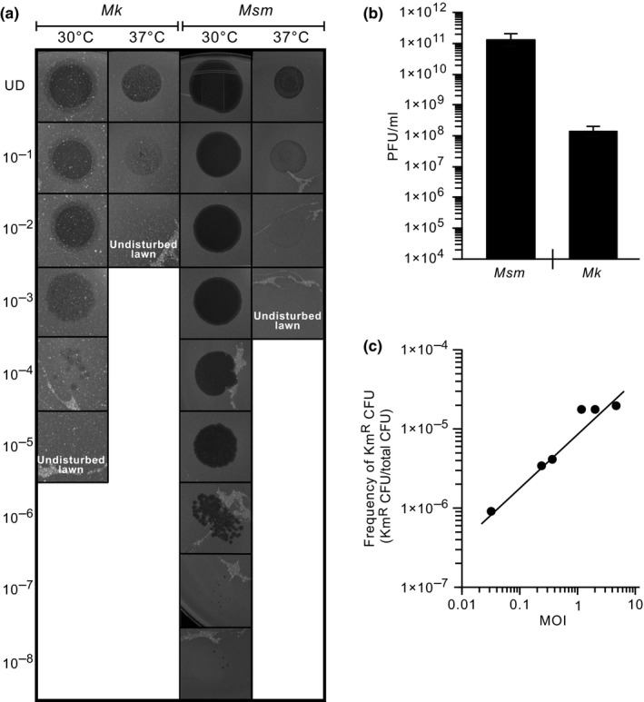
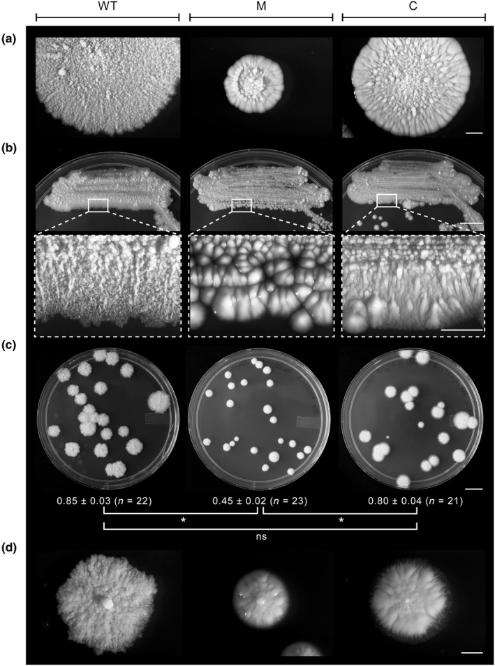
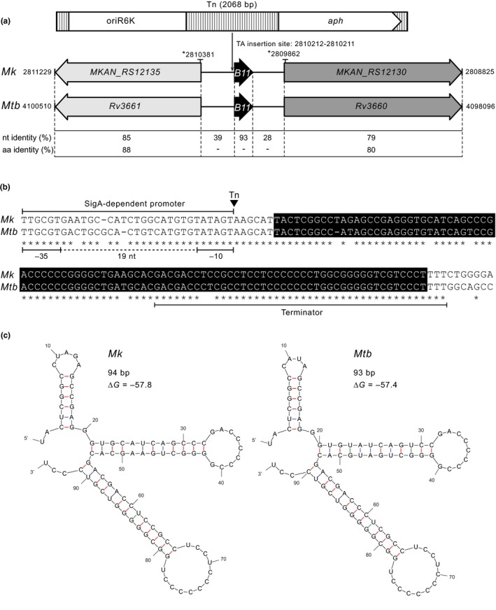
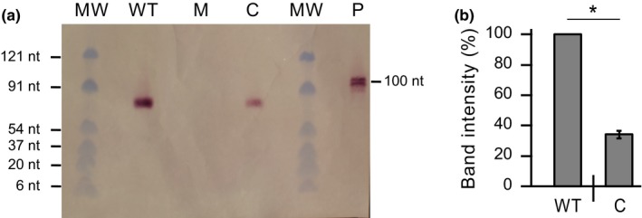
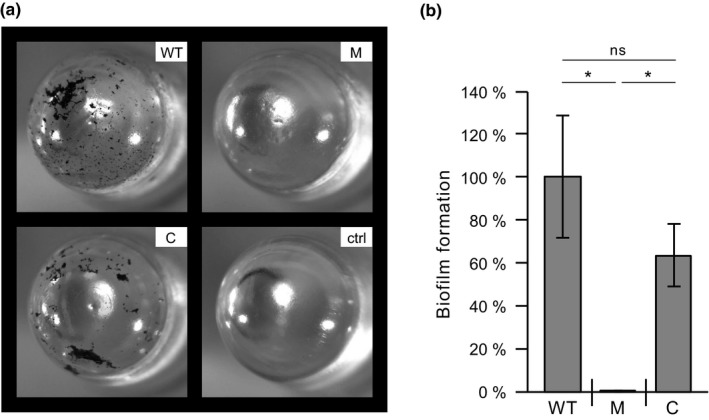
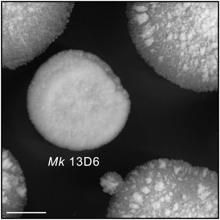
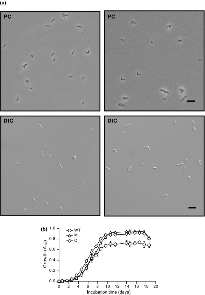
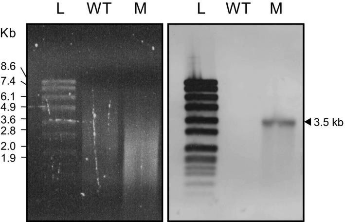
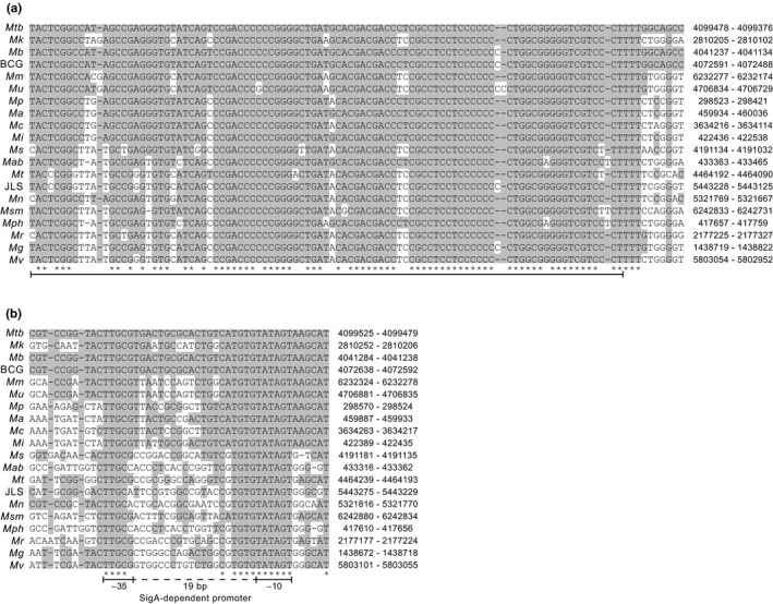
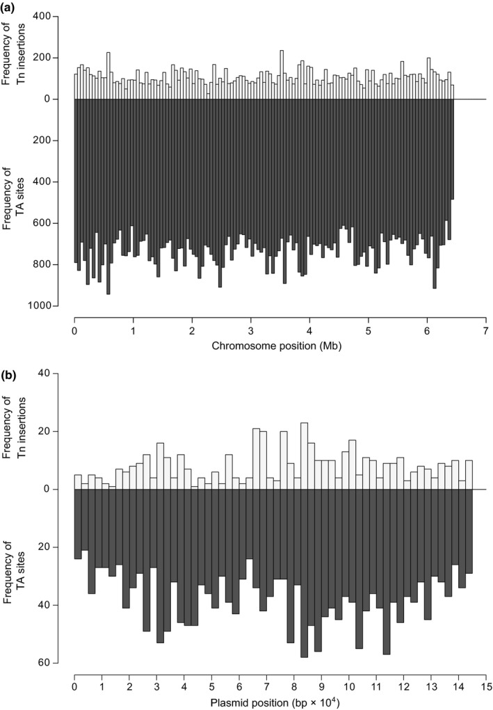
Mycobacterium kansasii (Mk) is a resilient opportunistic human pathogen that causes tuberculosis-like chronic pulmonary disease and mortality stemming from comorbidities and treatment failure. The standard treatment of Mk infections requires costly, long-term, multidrug courses with adverse side effects. The emergence of drug-resistant isolates further complicates the already challenging drug therapy regimens and threatens to compromise the future control of Mk infections. Despite the increasingly recognized global burden of Mk infections, the biology of this opportunistic pathogen remains essentially unexplored. In particular, studies reporting gene function or generation of defined mutants are scarce. Moreover, no transposon (Tn) mutagenesis tool has been validated for use in Mk, a situation limiting the repertoire of genetic approaches available to accelerate the dissection of gene function and the generation of gene knockout mutants in this poorly characterized pathogen. In this study, we validated the functionality of a powerful Tn mutagenesis tool in Mk and used this tool in conjunction with a forward genetic screen to establish a previously unrecognized role of a conserved mycobacterial small RNA gene of unknown function in colony morphology features and biofilm formation. We also combined Tn mutagenesis with next-generation sequencing to identify 12,071 Tn insertions that do not compromise viability in vitro. Finally, we demonstrated the susceptibility of the Galleria mellonella larva to Mk, setting the stage for further exploration of this simple and economical infection model system to the study of this pathogen.

Keywords: Galleria mellonella; Mycobacterium kansasii; biofilm; gene essentiality; nontuberculous mycobacteria; small noncoding RNA.


Conflict of interest statement

None declared.

Figures

Figure 1
Figure 1
Functionality of the ϕMycoMarT7‐based transposon mutagenesis system in Mycobacterium kansasii. (a) Images showing the plaque formation capacity of the ϕMycoMarT7 phage on lawns of Mk and Msm at the permissive (30°C) and nonpermissive (37°C) temperatures. The numbers to the left of the image represent a 10‐fold dilution series of the phage stock. UD, undiluted phage stock. (b) Comparison of plaque formation efficiency on lawns of Mk and Msm at the permissive temperature. The data represent mean ± SE of at least two PFU titrations. (c) Effect of the multiplicity of infection (MOI) used in the transduction mixture on the efficiency of generation of Mk Tn mutants
Figure 2
Figure 2
Morphology phenotypes of Mycobacterium kansasii strains. (a) Representative spot inoculation‐derived macrocolonies showing morphological features. Scale bar = 2 mm. (b) Plates with bacterial streaks (top; scale bar = 1 cm) and enlargements (bottom; scale bar = 2 mm) of the boxed sections showing surface topology and appearance features. (c) Plates with isolated single colonies. Scale bar = 1 cm. Colony diameter (cm) information is presented below each plate. The values shown represent mean ± SE. *Student's t test p values <.05; ns, no significant difference (p ≥ .05). (d) Representative single colonies showing morphology detail. Scale bar = 2 mm. WT, wt‐v; M, 13D6‐v; C, 13D6‐c
Figure 3
Figure 3
Mycobacterium kansasii 13D6 has a transposon insertion located 6 bp upstream of an sRNA gene (Mk B11) orthologous to the Mycobacterium tuberculosis sRNA gene B11. (a) Outline depicting the Tn (Escherichia coli replication origin oriR6k and aph KmR marker are represented), the location of the TA insertion site (chromosomal coordinates are shown), and the conservation of genetic features nearby the insertion site in the Mk chromosome (NC_022663.1) and the orthologous segment in the Mtb chromosome (NC_000962.3). Chromosomal coordinates of the segments are indicated. The coordinates of the chromosomal fragment included in the complementation plasmid pML‐B11 are marked by an asterisk (*). Nucleotide (nt) and amino acid (aa) identity between specific sections of the segments is provided. (b) Sequence alignment of Mk and Mtb B11 loci. The boxed sequences correspond to the annotated Mtb B11 gene. The location of the Tn (inverted triangle) and putative promoter and terminator sequences are depicted. (c) Predicted secondary structures for the sRNA products of the B11 genes. The minimum free energy associated with each structure is shown
Figure 4
Figure 4
Northern blot analysis of Mycobacterium kansasii B11 sRNA in M. kansasii strains. (a) Representative northern blot demonstrating the presence of B11 sRNA in the WT strain (wt‐v, lane WT) and complemented strain (13D6‐c, lane C) and its absence in the mutant strain (13D6‐v, lane M). Lane MW, molecular weight markers; lane P, B11 probe fragment (100 bp). (b) Northern blot‐based comparison of B11 expression in the WT (WT) and complemented (C) strains. The bands on the blot were quantified by densitometry analysis using ImageJ software (NIH, available in the public domain). The data represent mean ± SE of quantifications from three northern blots. *Student's t test p values <.0001
Figure 5
Figure 5
RT‐qPCR expression analysis of B11 and adjacent genes in Mycobacterium kansasii strains. (a) Expression of B11 in macrocolonies. (b) Expression of B11 in planktonic cells. (c) Expression of B11 gene neighbors in macrocolonies. (d) Expression of B11 gene neighbors in planktonic cells. WT, wt‐v; M, 13D6‐v; C, 13D6‐c. In‐bar values reflect fold reduction in the indicated strain relative to WT. Data represent mean ± SE of at least two independent experiments. Student's t test p values: *<.05; **<.01; ns, no significant difference (p ≥ .05)
Figure 6
Figure 6
Biofilm formation by Mycobacterium kansasii strains. (a) Representative image of crystal violet‐stained MBEC™ device pegs. (b) Quantification of biofilm formation on pegs using the crystal violet‐based colorimetric assay. WT, wt‐v; M, 13D6‐v; C, 13D6‐c; ctrl, sterile medium control. Data represent mean ± SE of three independent experiments. *Student's t test p values <.05; ns, no significant difference (p ≥ .05)
Figure 7
Figure 7
Kaplan–Meier survival curves demonstrating temperature‐dependent killing of Galleria mellonella larvae by Mycobacterium kansasii. WT, WT strain; M, 13D6 mutant; BC, PBS buffer control; NIC, noninjected control. Statistical differences between survival curves were assessed using the Log‐rank test. p values: ***<.0001; **.0053; *.0362; ns, no significant difference (p ≥ .05)
Figure A1
Figure A1
Section of the original screen plate showing the abnormal macrocolony morphology phenotype of Mycobacterium kansasii 13D6. Scale bar = 2 mm
Figure A2
Figure A2
The transposon insertion in Mycobacterium kansasii 13D6 does not compromise planktonic growth, nor does it affect cell morphology. (a) Representative phase contrast (PC) and differential interference contrast (DIC) microscopy images showing cells from exponentially growing cultures of the WT (left) and mutant (right) strains. Scale bars = 5 µm. Images were acquired using a Nikon Eclipse Ti inverted microscope (Nikon Instruments Inc.) (b) Growth curves of Mk strains. WT, wt‐v; M, 13D6‐v; and C, 13D6‐c. The growth curve data were generated in a multiwell plate‐based growth assay as described in Section 2. Values of each time point represent mean ± SE of three independent growth curves
Figure A3
Figure A3
Southern blot hybridization analysis of Mycobacterium kansasii 13D6. Agarose gel electrophoresis of SacII‐digested genomic DNA (left panel) and southern blot membrane after hybridization with a Tn‐specific probe (right panel). Lanes: L, DNA molecular weight ladder; WT, WT strain; M, 13D6 mutant
Figure A4
Figure A4
Conservation of the B11 gene locus in mycobacteria. (a) Sequence alignment of Mycobacterium tuberculosis B11, Mycobacterium kansasii B11, and predicted orthologs in other mycobacteria. (b) Sequence alignment of B11 promoter regions. Gray shading highlights identity with the Mtb sequence. Asterisks below the alignment indicate 100% conservation. The bar under the alignment of B11 orthologs marks the sequence annotated for Mtb B11. Abbreviations to the left and chromosomal coordinates to the right of the sequences correspond to the following bacteria and NCBI reference sequences, respectively. Mtb: M. tuberculosis strain H37Rv, NC_000962.3; Mk: M. kansasii strain ATCC 12478, NC_022663.1; Mb: Mycobacterium bovis strain AF2122/97, NC_002945; BCG: M. bovis BCG Pasteur 1173P2, NC_008769.1; Mm: Mycobacterium marinum strain M, NC_010612.1; Mu: Mycobacterium ulcerans strain Agy99, NC_008611.1; Mp: Mycobacterium parascrofulaceum strain ATCC BAA‐614 SCAFFOLD1, NZ_GG770553.1; Ma: Mycobacterium avium strain 104, NC_008595.1; Mc: Mycobacterium columbiense strain CECT 3035, NZ_CP020821.1; Mi: Mycobacterium intracellulare strain ATCC 13950, NC_016946.1; Ms: Mycobacterium sinense strain JDM601, NC_015576.1; Mab: Mycobacterium abscessus strain ATCC 19977, NC_010397.1; Mt: Mycobacterium thermoresistible strain NCTC10409, NZ_LT906483.1; JLS: M. sp. JLS, NC_009077.1; Mn: Mycobacterium neoaurum strain VKM Ac‐1815D, NC_023036.2; Msm: Mycobacterium smegmatis strain mc2155, NC_018289.1; Mph: Mycobacterium phlei strain NCTC8151, NZ_LR134347.1; Mr: Mycobacterium rhodesiae strain NBB3, NC_016604.1; Mg: Mycobacterium gilvium strain PYR‐GCK, NC_009338.1; and Mv: Mycobacterium vanbaalenii strain PYR‐1, NC_008726.1
Figure A5
Figure A5
Mycobacteriophage L5 integration attB site in Mycobacterium kansasii. Alignment of the 62‐bp attB site in Mycobacterium smegmatis strain mc2155 (Msm, NCBI reference sequence: NC_008596.1) and the predicted attB site in M. kansasii (Mk, NCBI reference sequence: NC_022663.1). Longer bar, 43‐bp attB common core. Shorter bar, 29‐bp necessary and sufficient for attB function. Chromosomal coordinates are indicated
Figure A6
Figure A6
Histograms showing overviews of frequency distributions of Tn insertions and TA dinucleotide sites along the chromosome (a) and the plasmid pMK12478 (b) of Mycobacterium kansasii. The data of the Tn insertions for the chromosome and plasmid are derived from Tables S1 and S2, respectively. The data of TA dinucleotide sites for the chromosome and plasmid are derived from NCBI Reference Sequences NC_022663.1 and NC_022654.1, respectively. The data presented for the chromosome and plasmid are organized in bins of 50,000 and 2,500 bp, respectively.

References

    1. Aldridge, B. B. , Keren, I. , & Fortune, S. M. (2014). The spectrum of drug susceptibility in mycobacteria. Microbiology Spectrum, 2(5), MGM2-0031-2013. 10.1128/microbiolspec.MGM2-0031-2013 - DOI - PubMed
    1. Arnvig, K. B. , Cortes, T. , & Young, D. B. (2014). Noncoding RNA in mycobacteria. Microbiology Spectrum, 2(2), MGM2-0029-2013. 10.1128/microbiolspec.MGM2-0029-2013 - DOI - PubMed
    1. Arnvig, K. B. , & Young, D. B. (2009). Identification of small RNAs in Mycobacterium tuberculosis . Molecular Microbiology, 73(3), 397–408. 10.1111/j.1365-2958.2009.06777.x - DOI - PMC - PubMed
    1. Bakula, Z. , Safianowska, A. , Nowacka‐Mazurek, M. , Bielecki, J. , & Jagielski, T. (2013). Short communication: Subtyping of Mycobacterium kansasii by PCR‐restriction enzyme analysis of the hsp65 gene. BioMed Research International, 2013, 178725 10.1155/2013/178725 - DOI - PMC - PubMed
    1. Bardarov, S. , Kriakov, J. , Carriere, C. , Yu, S. , Vaamonde, C. , McAdam, R. A. , … Jacobs, W. R. (1997). Conditionally replicating mycobacteriophages: A system for transposon delivery to Mycobacterium tuberculosis . Proceedings of the National Academy of Sciences of the United States of America, 94(20), 10961–10966. 10.1073/pnas.94.20.10961 - DOI - PMC - PubMed

Publication types

MeSH terms

LinkOut - more resources