

CD73 or CD39 Deletion Reveals Different Mechanisms of Formation for Spontaneous and Mechanically Stimulated Adenosine and Sex Specific Compensations in ATP Degradation
- PMID: 32083837
- PMCID: PMC7335217
- DOI: 10.1021/acschemneuro.9b00620
CD73 or CD39 Deletion Reveals Different Mechanisms of Formation for Spontaneous and Mechanically Stimulated Adenosine and Sex Specific Compensations in ATP Degradation
Abstract
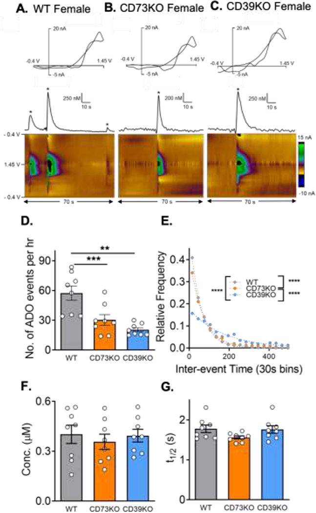
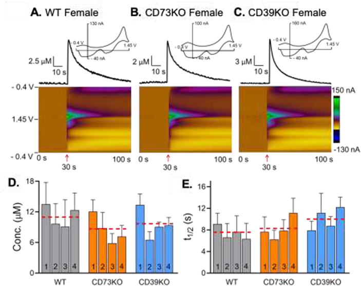
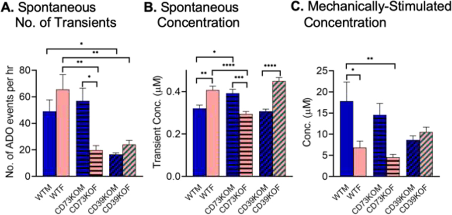
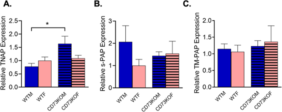
Adenosine is important for local neuromodulation, and rapid adenosine signaling can occur spontaneously or after mechanical stimulation, but little is known about how adenosine is formed in the extracellular space for those stimulations. Here, we studied mechanically stimulated and spontaneous adenosine to determine if rapid adenosine is formed by extracellular breakdown of adenosine triphosphate (ATP) using mice globally deficient in extracellular breakdown enzymes, either CD39 (nucleoside triphosphate diphosphohydrolase 1, NTPDase1) or CD73 (ecto-5'-nucleotidase). CD39 knockout (KO) mice have a lower frequency of spontaneous adenosine events than wild-type (WT, C57BL/6). Surprisingly, CD73KO mice demonstrate sex differences in spontaneous adenosine; males maintain similar event frequencies as WT, but females have significantly fewer events and lower concentrations. Examining the mRNA expression of other enzymes that metabolize ATP revealed tissue nonspecific alkaline phosphatase (TNAP) was upregulated in male CD73KO mice, but not secreted prostatic acid phosphatase (PAP) or transmembrane PAP. Thus, TNAP upregulation compensates for CD73 loss in males but not in females. These sex differences highlight that spontaneous adenosine is formed by metabolism of extracellular ATP by many enzymes. For mechanically stimulated adenosine, CD39KO or CD73KO did not change stimulation frequency, concentration, or t1/2. Thus, the mechanism of formation for mechanically stimulated adenosine is likely direct release of adenosine, different than spontaneous adenosine. Understanding these different mechanisms of rapid adenosine formation will help to develop pharmacological treatments that differentially target modes of rapid adenosine signaling, and all treatments should be studied in both sexes, given possible differences in extracellular ATP degradation.
Keywords: Adenosine; CD39KO; CD73KO; PAP; TNAP; hippocampus; in vivo; mechanosensitive; sex differences; spontaneous adenosine.
Figures
Similar articles
-
Extracellular generation of adenosine by the ectonucleotidases CD39 and CD73 promotes dermal fibrosis.Am J Pathol. 2013 Dec;183(6):1740-1746. doi: 10.1016/j.ajpath.2013.08.024. Am J Pathol. 2013. PMID: 24266925 Free PMC article.
-
The adenosine generating enzymes CD39/CD73 control microglial processes ramification in the mouse brain.PLoS One. 2017 Apr 4;12(4):e0175012. doi: 10.1371/journal.pone.0175012. eCollection 2017. PLoS One. 2017. PMID: 28376099 Free PMC article.
-
CD73 controls ocular adenosine levels and protects retina from light-induced phototoxicity.Cell Mol Life Sci. 2022 Feb 25;79(3):152. doi: 10.1007/s00018-022-04187-4. Cell Mol Life Sci. 2022. PMID: 35212809 Free PMC article.
-
The role of the CD39-CD73-adenosine pathway in liver disease.J Cell Physiol. 2021 Feb;236(2):851-862. doi: 10.1002/jcp.29932. Epub 2020 Jul 10. J Cell Physiol. 2021. PMID: 32648591 Review.
-
Extracellular adenosine generation in the regulation of pro-inflammatory responses and pathogen colonization.Biomolecules. 2015 May 5;5(2):775-92. doi: 10.3390/biom5020775. Biomolecules. 2015. PMID: 25950510 Free PMC article. Review.
Cited by
-
Dual-Channel Electrochemical Measurements Reveal Rapid Adenosine is Localized in Brain Slices.ACS Chem Neurosci. 2022 Feb 16;13(4):477-485. doi: 10.1021/acschemneuro.1c00679. Epub 2022 Jan 25. ACS Chem Neurosci. 2022. PMID: 35077156 Free PMC article.
-
Purinergic System Transcript Changes in the Dorsolateral Prefrontal Cortex in Suicide and Major Depressive Disorder.Int J Mol Sci. 2025 Feb 20;26(5):1826. doi: 10.3390/ijms26051826. Int J Mol Sci. 2025. PMID: 40076453 Free PMC article.
-
Adenosine signaling: Optimal target for gastric cancer immunotherapy.Front Immunol. 2022 Sep 16;13:1027838. doi: 10.3389/fimmu.2022.1027838. eCollection 2022. Front Immunol. 2022. PMID: 36189223 Free PMC article. Review.
-
Spontaneous, transient adenosine release is not enhanced in the CA1 region of hippocampus during severe ischemia models.J Neurochem. 2021 Dec;159(5):887-900. doi: 10.1111/jnc.15496. Epub 2021 Sep 20. J Neurochem. 2021. PMID: 34453336 Free PMC article.
-
The elegant complexity of mammalian ecto-5'-nucleotidase (CD73).Trends Cell Biol. 2021 Oct;31(10):829-842. doi: 10.1016/j.tcb.2021.05.008. Epub 2021 Jun 8. Trends Cell Biol. 2021. PMID: 34116887 Free PMC article. Review.
References
Publication types
MeSH terms
Substances
Grants and funding
LinkOut - more resources
Full Text Sources
Research Materials
