. 2020 Feb 21;15(2):e0229376.
doi: 10.1371/journal.pone.0229376. eCollection 2020.

The acid-base-nucleophile catalytic triad in ABH-fold enzymes is coordinated by a set of structural elements

Affiliations

The acid-base-nucleophile catalytic triad in ABH-fold enzymes is coordinated by a set of structural elements

Alexander Denesyuk et al. PLoS One. .

Abstract

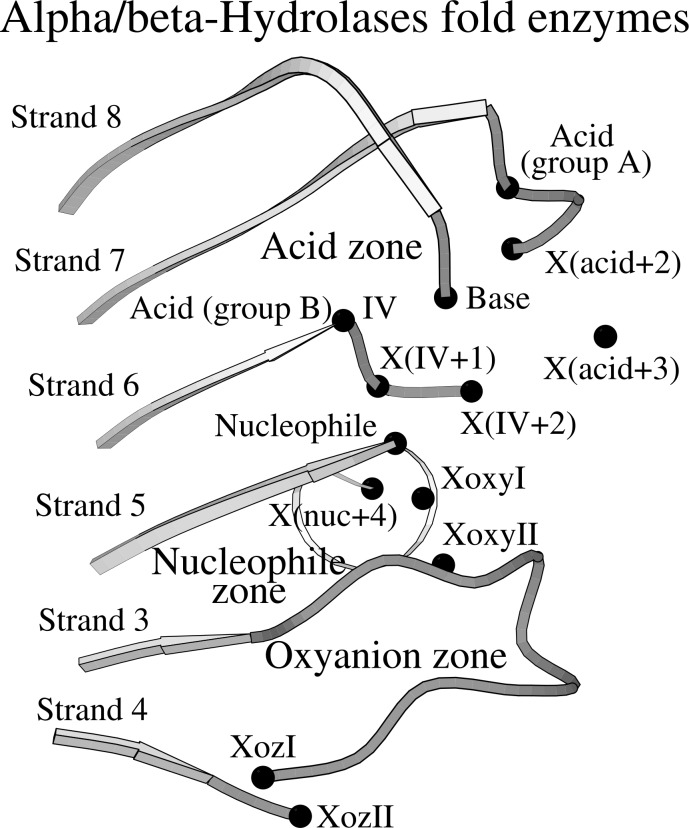
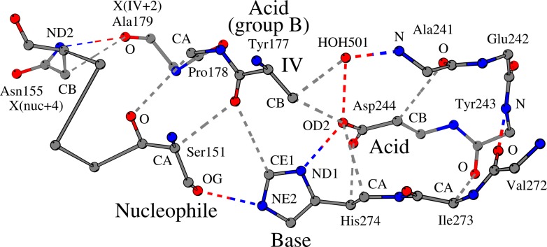
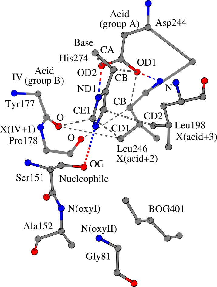
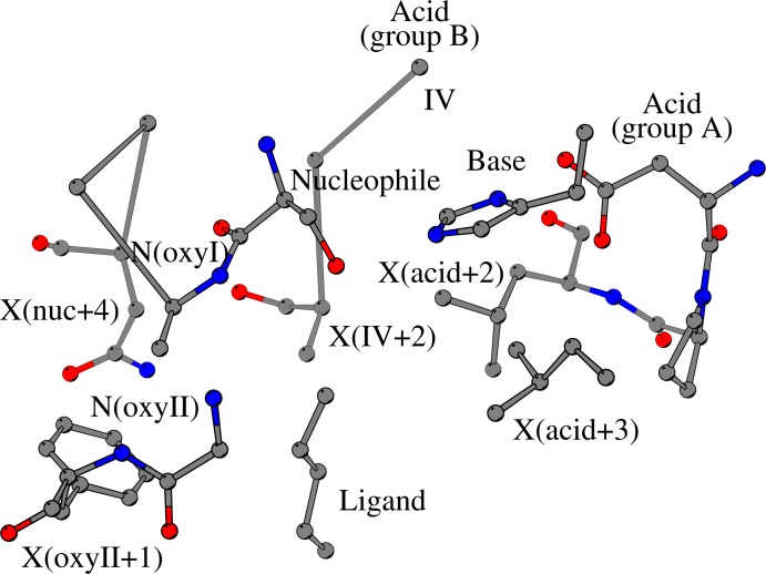
The alpha/beta-Hydrolases (ABH) are a structural class of proteins that are found widespread in nature and includes enzymes that can catalyze various reactions in different substrates. The catalytic versatility of the ABH fold enzymes, which has been a valuable property in protein engineering applications, is based on a similar acid-base-nucleophile catalytic mechanism. In our research, we are concerned with the structure that surrounds the key units of the catalytic machinery, and we have previously found conserved structural organizations that coordinate the catalytic acid, the catalytic nucleophile and the residues of the oxyanion hole. Here, we explore the architecture that surrounds the catalytic histidine at the active sites of enzymes from 40 ABH fold families, where we have identified six conserved interactions that coordinate the catalytic histidine next to the catalytic acid and the catalytic nucleophile. Specifically, the catalytic nucleophile is coordinated next to the catalytic histidine by two weak hydrogen bonds, while the catalytic acid is directly involved in the coordination of the catalytic histidine through by two weak hydrogen bonds. The imidazole ring of the catalytic histidine is coordinated by a CH-π contact and a hydrophobic interaction. Moreover, the catalytic triad residues are connected with a residue that is located at the core of the active site of ABH fold, which is suggested to be the fourth member of a "structural catalytic tetrad". Besides their role in the stability of the catalytic mechanism, the conserved elements of the catalytic site are actively involved in ligand binding and affect other properties of the catalytic activity, such as substrate specificity, enantioselectivity, pH optimum and thermostability of ABH fold enzymes. These properties are regularly targeted in protein engineering applications, and thus, the identified conserved structural elements can serve as potential modification sites in order to develop ABH fold enzymes with altered activities.


Conflict of interest statement

The authors have declared that no competing interests exist.

Figures

Fig 1
Fig 1. Conserved structural motifs at the catalytic site of ABH fold enzymes.
Most key features of the catalytic machinery are coordinated by conserved structural organizations within the same plane as the central β-sheet of the ABH fold. Specifically, the catalytic acid (“Acid”), which is located at the turn that follows strand β7 (“group A”) or at the C-terminus of strand β6 (“group B”, position “IV”), is coordinated by the structural organization that is called “Catalytic acid zone” (“Acid zone”), while the catalytic nucleophile (“Nucleophile”) and the two residues that help form the oxyanion hole (“XoxyI” and “XoxyII”) are coordinated by the overlapping “Nucleophile zone” and “Oxyanion zone”. The catalytic histidine (“Base”) is located at a flexible loop that follows strand β8 and is linked with the β-sheet at the C-terminus of strand β6. The residues of the catalytic machinery, two conserved residues of Oxyanion zone (“XozI” and “XozII”), four residues of catalytic acid loops (“X(acid+2)”, “X(acid+3)”, “X(IV+1)” and “X(IV+2)”) and one residue of the nucleophile elbow (“X(nuc+4)”) are indicated with black dots.
Fig 2
Fig 2. Coordination of the catalytic nucleophile-histidine pair and the catalytic histidine-acid pair in the carboxylesterase SshEstI (PDB ID:3WJ1).
The catalytic nucleophile (“Nucleophile”, Ser151 in SshEstI) is hydrogen bonded (OG/Ser151 –NE2/His274) to the catalytic histidine (“Base”, His274 in SshEstI) as part of the standard interaction network of the residues of the catalytic machinery. However, three additional conserved interactions ensure the fine turning of the two catalytic residues next to each other: The main-chain oxygen atom of Tyr177 that is located at the C-terminus of strand β6 (termed and shown as “IV”) forms two weak hydrogen bonds: one with the catalytic nucleophile (O/Tyr177 –CA/Ser151) and another with the catalytic histidine (O/Tyr177 –CE1/His274); a third contact (O/Ser151 –CA/Pro178) is formed between the catalytic nucleophile and Pro178 (located ahead of position IV) and coordinates the catalytic nucleophile. Thus, these three interactions support the optimal arrangement of the catalytic nucleophile-histidine pair. The catalytic histidine (His274) interacts (ND1/His274 –OD2/Asp244) with the catalytic acid (“Acid”, Asp244 in SshEstI) and is further supported by two weak hydrogen bonds: OD2/Asp244 –CA/His274 and OD1/Asp244 –CB/His274. Interactions of the catalytic acid zone that are associated with the coordination of the catalytic histidine and other contacts located nearby the catalytic site are shown. Gray dashed lines, weak hydrogen bonds; colored dashed lines, standard hydrogen bonds.
Fig 3
Fig 3. Coordination of the imidazole ring of the catalytic histidine in the carboxylesterase SshEstI (PDB ID:3WJ1).
The optimal arrangement of catalytic residues requires the proper positioning of the imidazole ring of the catalytic histidine (“Base”, His274 in SshEstI) relative to the side chains of the catalytic nucleophile (“Nucleophile”, Ser151 in SshEstI) and the catalytic acid (“Acid”, Asp244 in SshEstI). The catalytic histidine is anchored to the β-sheet through its contact with Tyr177 (position IV) and is directly coordinated by the catalytic acid Asp244. The imidazole ring of the catalytic histidine is coordinated by two interactions: a hydrophobic interaction (CD1/Leu246 –CE1/His274) with Leu246 (X(acid+2)), which is located two sequence positions after the catalytic acid, and a CH–π interaction (CD2/Leu198 –π/His274) with Leu198 (X(acid+3)). The residues at the C-terminal end of strand β6, Tyr177 (IV) and Pro178 (IV+1), also interact with Leu246, which is situated at the turn that accommodates the catalytic acid. The residues, “OxyI” and “OxyII”, which help form the oxyanion hole, and the bound ligand “BOG401” of carboxylesterase SshEstI are indicated. Gray dashed lines represent weak hydrogen bonds; colored dashed lines, standard hydrogen bonds.
Fig 4
Fig 4. Structural elements at the active site of the ABH fold enzymes surround the bound ligand.
Recognized substrate molecules (shown as “Ligand”) are bound to the active sites of ABH fold enzymes in order to be hydrolyzed. Key residues of the catalytic machinery, such as the catalytic histidine (“Base”), the catalytic nucleophile (“Nucleophile”) and the residues that help form the oxyanion hole (“oxyI” and “oxyII”) often interact with the bound substrate; the catalytic acid, either at its canonical position (at the turn that follows strand β7, “Acid (group A)”) or at its alternate position (at the C-terminal end of strand β6, “Acid (group B)”) does not participate in ligand binding. Several residues, which belong to the structural core of ABH fold enzymes, participate both in the coordination of the residues of the catalytic machinery and in ligand binding, including the residues that are located two and three sequence positions after the catalytic acid, illustrated as “X(acid+2)” and “X(acid+3)” respectively, and the residue at the C-terminus of strand β6. Other residues, usually comprising the substrate binding core, are illustrated as “X(IV+2)” for the residue that is located two positions after the C-terminus of strand β6, “X(oxyII+1)” for the residue that follows the second residue that helps form the oxyanion hole, and “X(nuc+4)” for the residue that is located four positions after the catalytic nucleophile.

References

    1. Nardini M, Dijkstra BW (1999) α/β-Hydrolase fold enzymes: the family keeps growing. Curr Opin Struct Biol 9:732–737. 10.1016/s0959-440x(99)00037-8 - DOI - PubMed
    1. Mindrebo JT, Nartey CM, Seto Y, Burkart MK, Noel JP (2016) Unveiling the functional diversity of the alpha/beta-hydrolase superfamily in the plant kingdom. Curr Opin Struct Biol 41:233–246. 10.1016/j.sbi.2016.08.005 - DOI - PMC - PubMed
    1. Lenfant N, Hotelier T, Velluet E, Bourne Y, Marchot P, Chatonnet A (2013) ESTHER, the database of the α/β-hydrolase fold superfamily of proteins: tools to explore diversity of functions. Nucleic Acids Res 41:D423–D429. 10.1093/nar/gks1154 - DOI - PMC - PubMed
    1. Jochens H, Hesseler M, Stiba K, Padhi SK, Kazlauskas RJ, Bornscheuer UT (2011) Protein engineering of α/β-hydrolase fold enzymes. ChemBioChem 12:1508–1517. 10.1002/cbic.201000771 - DOI - PubMed
    1. Rauwerdink A, Kazlauskas RJ (2015) How the same core catalytic machinery catalyzes 17 different reactions: the serine-histidine-aspartate catalytic triad of α/β-hydrolase fold enzymes. ACS Catalysis 5:6153–6176. 10.1021/acscatal.5b01539 - DOI - PMC - PubMed

Publication types