

Long-term Evolution and Remodeling of Soft Drusen in Rhesus Macaques
- PMID: 32084273
- PMCID: PMC7326602
- DOI: 10.1167/iovs.61.2.32
Long-term Evolution and Remodeling of Soft Drusen in Rhesus Macaques
Abstract
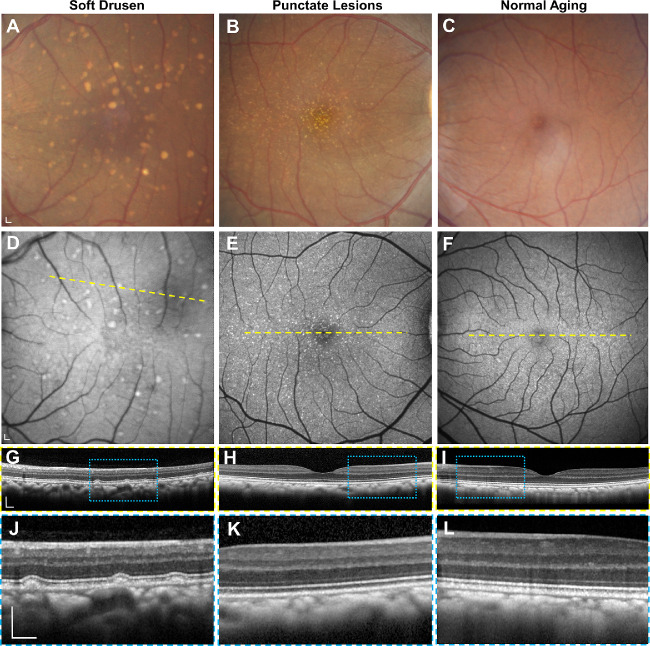
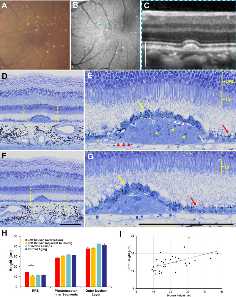
Purpose: To characterize the evolution and structure of soft drusen in aged rhesus macaques using in vivo multimodal retinal imaging and ex vivo histologic and ultrastructural analyses as a nonhuman primate model of early age-related macular degeneration (AMD).
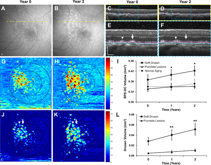
Methods: Multimodal imaging including fundus photography, spectral domain optical coherence tomography (SD-OCT), and fundus autofluorescence (FAF) were used to characterize and track individual drusen lesions in 20 aged rhesus macaques (mean age 23.3 ± 2.7 years) with drusenoid lesions over 2 years, followed by semithin histologic analysis and transmission electron microscopy (TEM).
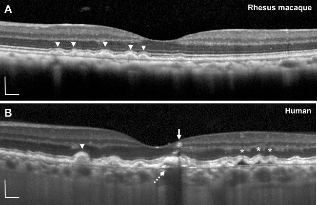
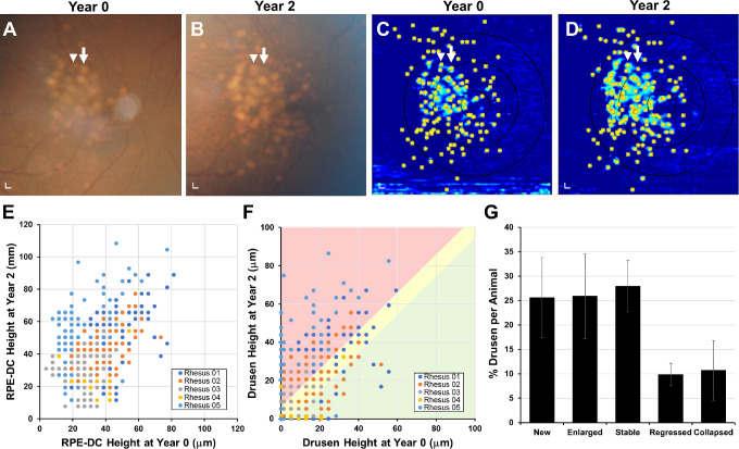
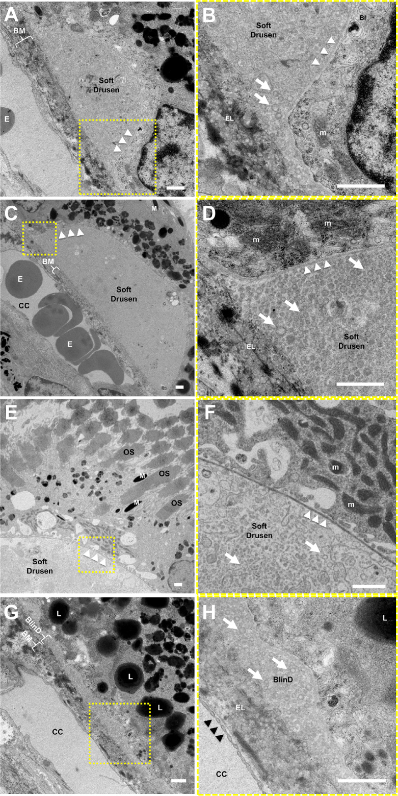
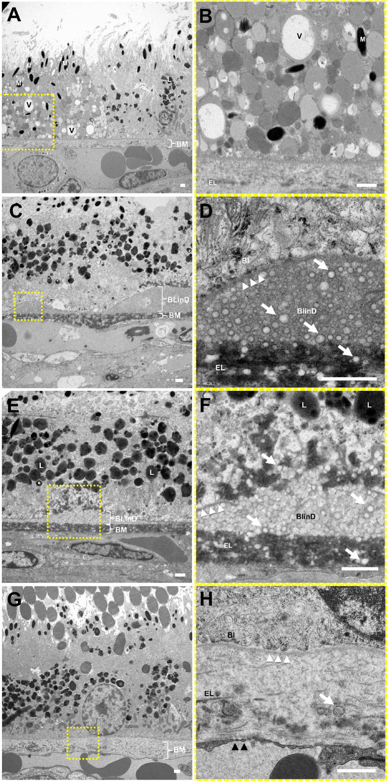
Results: Although most drusen gradually increased in size, a portion spontaneously regressed or collapsed over 2 years. Histologic analyses showed that soft drusen exhibit hypertrophy and dysmorphia of overlying retinal pigment epithelium (RPE), as seen in early and intermediate AMD, but do not exhibit RPE atrophy, RPE migration, or photoreceptor degeneration characteristic of advanced AMD. Ultrastructure of soft drusen showed abundant lipid particles within Bruch's membrane and AMD-related basal linear deposits (BlinD) resembling those in human drusen.
Conclusions: The dynamic remodeling, histologic findings, and ultrastructural features of soft drusen in aged rhesus macaques support nonhuman primates as an animal model of early AMD and reveal important insights into drusen biogenesis and AMD development.
Conflict of interest statement
Disclosure:
Figures
References
-
- Holz FG, Strauss EC, Schmitz-Valckenberg S, van Lookeren Campagne M. Geographic atrophy: clinical features and potential therapeutic approaches. Ophthalmology. 2014; 121: 1079–1091. - PubMed
-
- Age-Related Eye Disease Study Research Group. The Age-Related Eye Disease Study system for classifying age-related macular degeneration from stereoscopic color fundus photographs: the Age-Related Eye Disease Study Report Number 6. Am J Ophthalmol. 2001; 132: 668–681. - PubMed
