. 2020 May;190(5):1118-1136.
doi: 10.1016/j.ajpath.2020.01.004. Epub 2020 Feb 19.

Fibroblast Activation Protein Regulates Lesion Burden and the Fibroinflammatory Response in Apoe-Deficient Mice in a Sexually Dimorphic Manner

Affiliations

Fibroblast Activation Protein Regulates Lesion Burden and the Fibroinflammatory Response in Apoe-Deficient Mice in a Sexually Dimorphic Manner

James Monslow et al. Am J Pathol. 2020 May.

Abstract

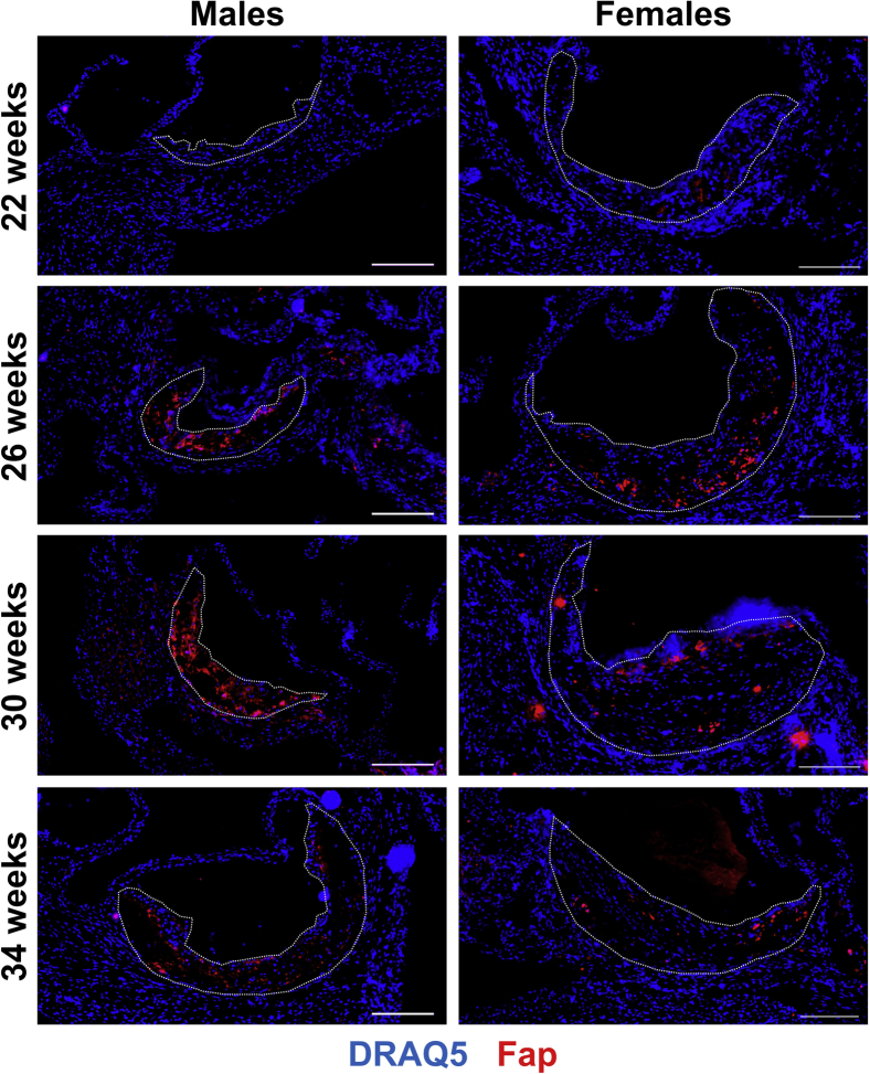
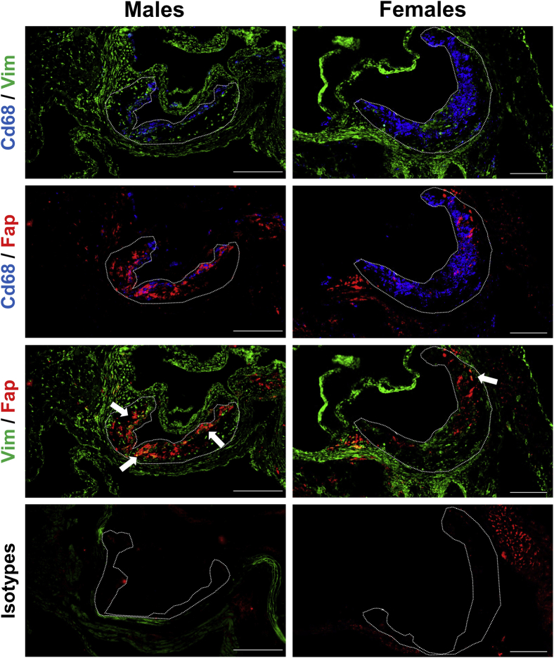
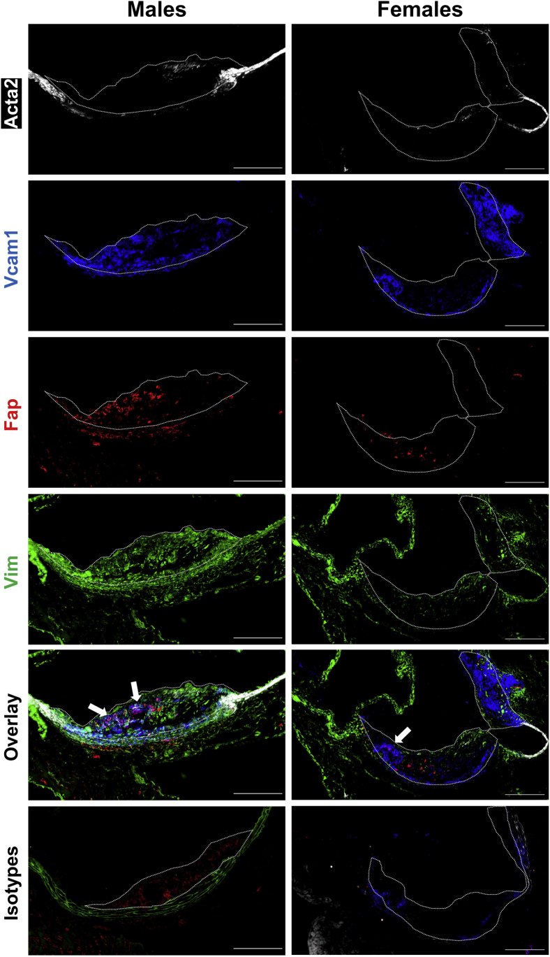
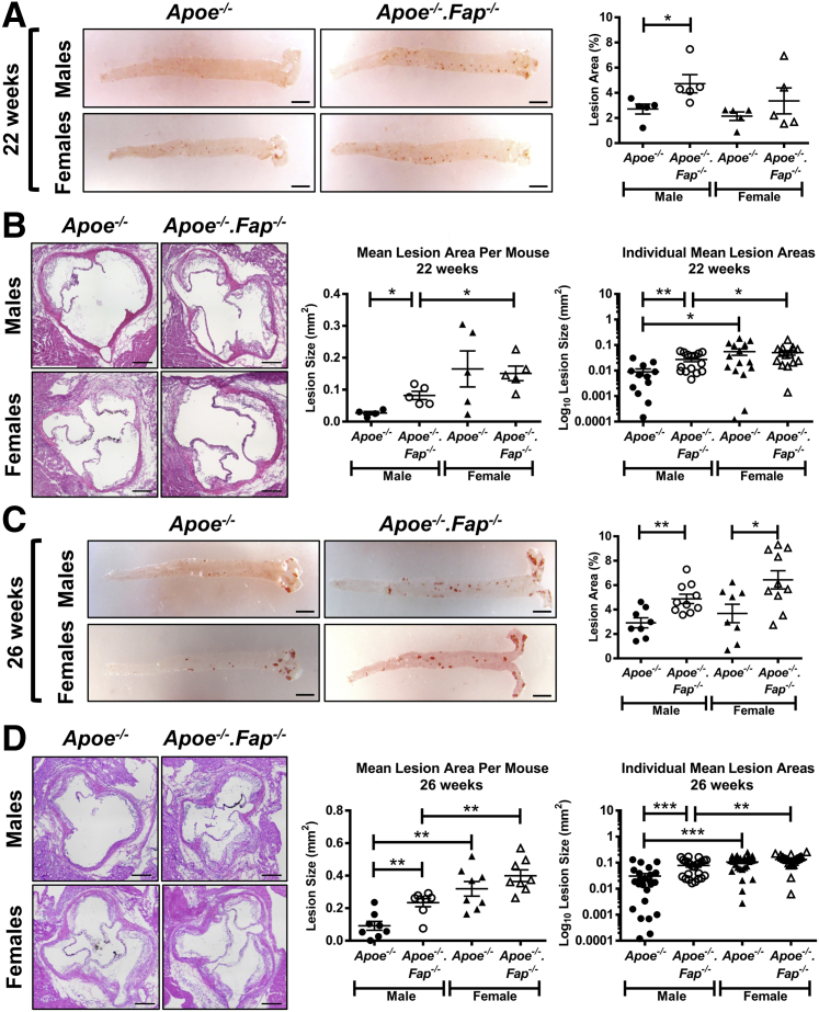
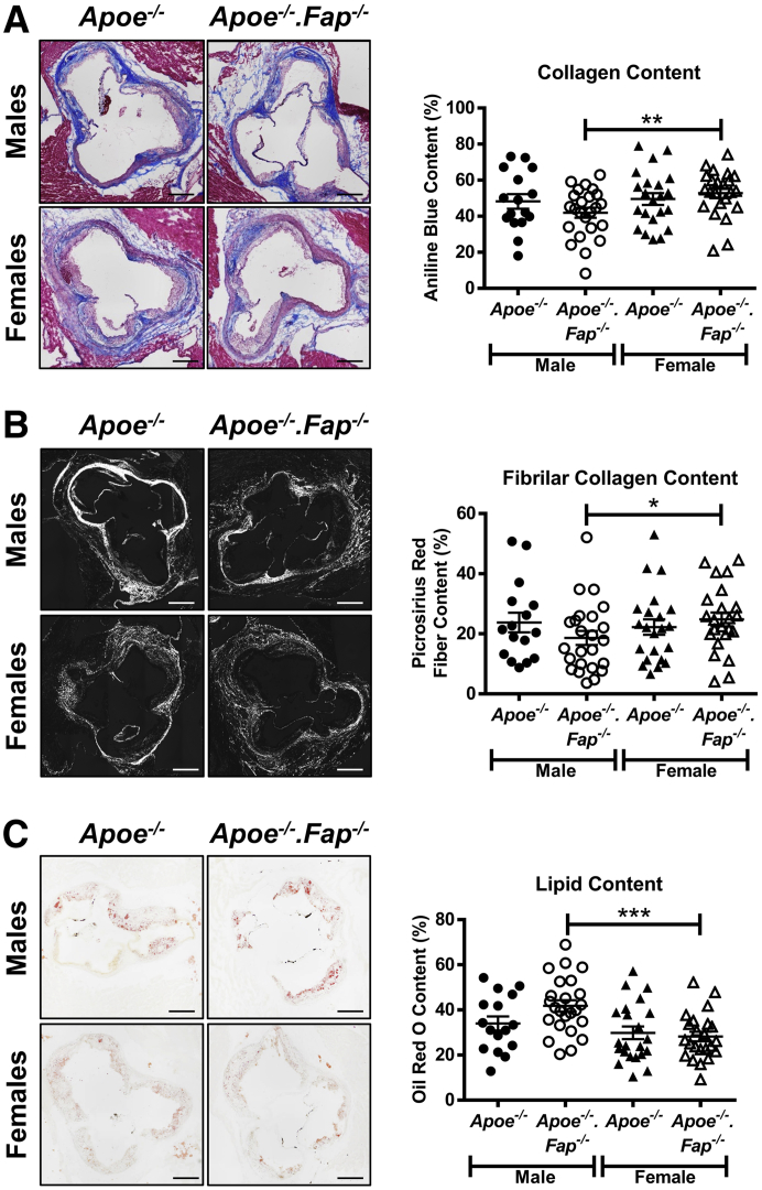
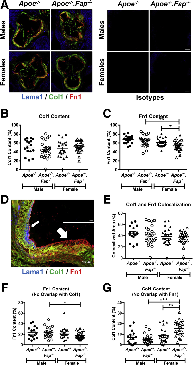
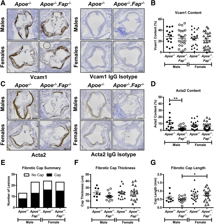
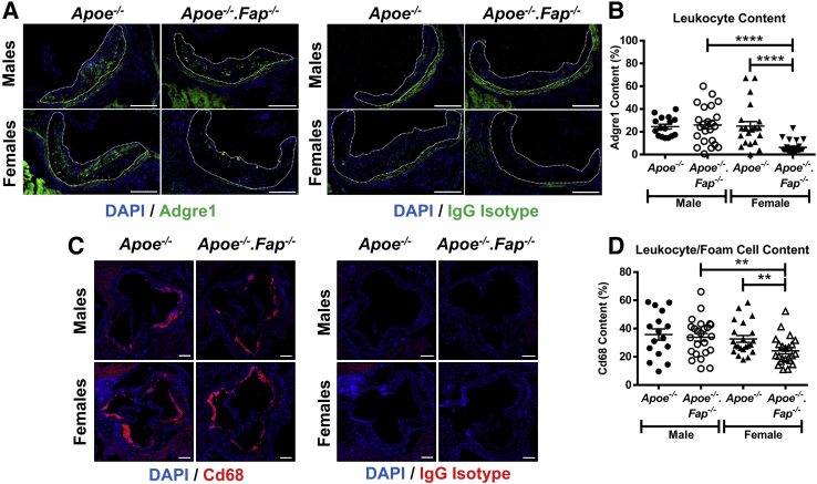
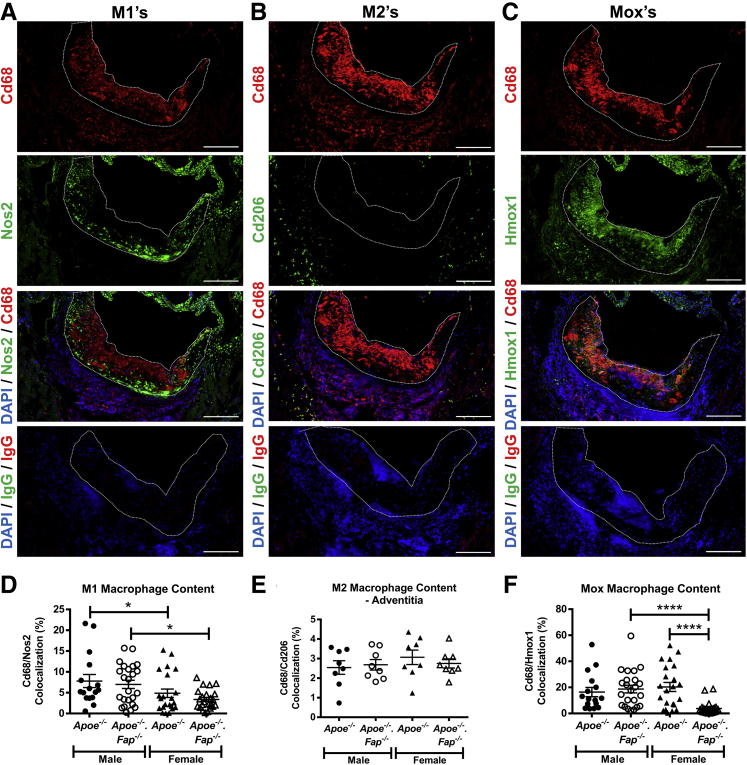
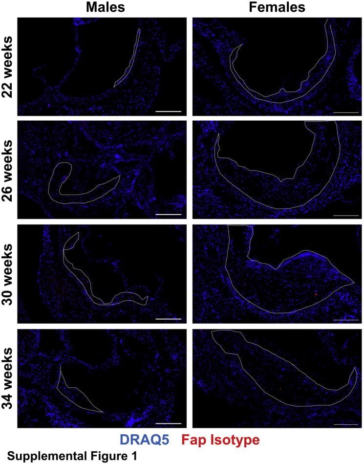
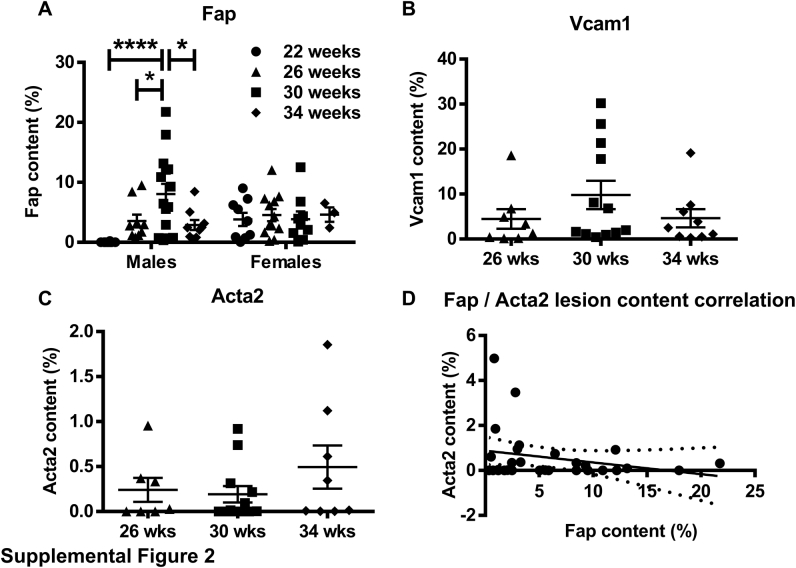
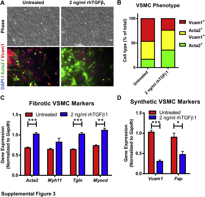
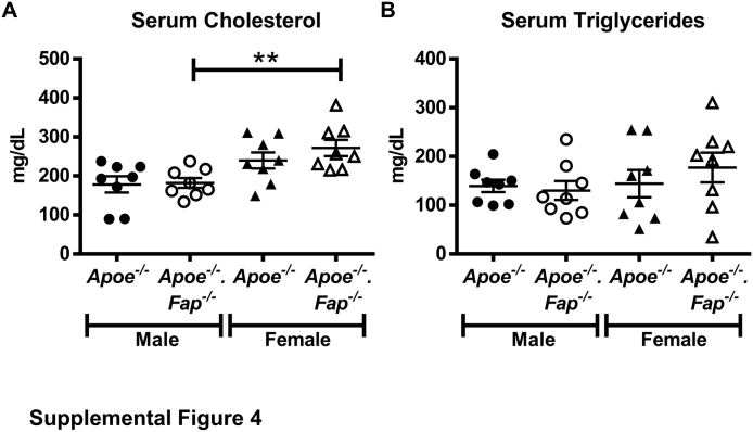
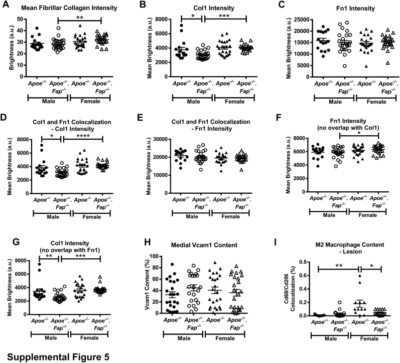
Fibroblast activation protein (FAP) has been established as an inducible and mesenchymal cell-specific mediator of disease progression in cancer and fibrosis. Atherosclerosis is a fibroinflammatory disease, and FAP was previously reported to be up-regulated in human atherosclerotic plaques compared with normal vessel. We investigated the spatial and temporal distribution of Fap-expressing cells in a murine model of atherosclerosis and used a genetic approach to determine if and how Fap affected disease progression. Fap was found to be expressed predominantly on vascular smooth muscle cells in lesions of athero-prone Apoe-/- mice. Global deletion of Fap (Fap-/-) in Apoe-/- mice accelerated atherosclerotic disease progression in both males and females, with the effect observed earlier in males. Sex-specific effects on lesion morphology were observed. Relative levels of extracellular matrix, fibrotic, and inflammatory cell content were comparable in lesions in male mice regardless of Fap status. In contrast, lesions in Fap-/- female mice were characterized by a more fibrotic composition due to a reduction in inflammation, specifically a reduction in Mox macrophages. Combined, these data suggest that Fap restrains the progression of atherosclerosis and may contribute to the sexually dimorphic susceptibility to atherosclerosis by regulating the balance between inflammation (an indicator of vulnerability to plaque rupture) and fibrosis (an indicator of plaque stability).


Figures

Figure 1
Figure 1
Spatiotemporal expression of fibroblast activation protein (Fap) in atherosclerotic lesions. Male and female Apoe−/− mice were maintained on a chow diet and aged to 22, 26, 30, or 34 weeks. Then 10-μm frozen sections of aortic roots were co-stained with antibodies targeted to Fap (red) and counterstained with DRAQ5 nuclear stain (blue). Atherosclerotic lesions are highlighted with dotted lines. Scale bars = 200 μm.
Figure 2
Figure 2
Fibroblast activation protein (Fap) expression is localized to mesenchymal cell–rich but not inflammatory cell–rich regions of atherosclerotic lesions. Male and female Apoe−/− mice were maintained on a chow diet and aged to 26 weeks. Then 10-μm frozen sections of aortic roots were co-stained with antibodies targeted to inflammatory regions [CD68 (blue)], mesenchymal regions [vimentin (Vim) (green)], and Fap (red) or isotype IgG controls. Atherosclerotic lesions are highlighted with dotted lines. Arrows indicate Fap/Vim overlapping regions (yellow). Scale bars = 200 μm.
Figure 3
Figure 3
Fibroblast activation protein (Fap) expression defines a subpopulation of mesenchymal cells. Male and female Apoe−/− mice were maintained on a chow diet and aged to 26 weeks. Multiplex staining of 10-μmol/L frozen sections of aortic roots was performed with antibodies targeted to mesenchymal vascular smooth muscle cells markers α-smooth muscle actin (Acta2) (white), vascular cell adhesion molecule 1 (Vcam1) (blue), Fap (red), and vimentin (Vim) (green). Atherosclerotic lesions are highlighted with dotted lines. Arrows highlight Fap/Vcam1 overlapping regions (magenta). Scale bars = 200 μm.
Figure 4
Figure 4
Deletion of fibroblast activation protein (Fap) accelerates atherosclerotic lesion progression in vivo. Male and female Apoe−/− and Apoe−/−.Fap−/− mice were maintained on a chow diet and aged to 22 or 26 weeks. A and C: Aortas were isolated and stained ex vivo with Oil Red O to visualize atherosclerotic lesions (red). Lesion burden was quantified as percentage of lesion area of the total aortic area. B and D: 10-μmol/L frozen cross-sections of aortic roots were stained with hematoxylin and eosin to visualize lesions. Lesion area was quantified and graphed as separate data points for each lesion or as mean lesion area per mouse. n = 4 to 5 mice (12 to 15 aortic root lesions) at 22 weeks and 8 to 10 mice (24 aortic root lesions) at 26 weeks. ∗P ≤ 0.05, ∗∗P ≤ 0.01, and ∗∗∗P ≤ 0.001. Scale bars: 2.5 mm (A and C); 200 μm (B and D).
Figure 5
Figure 5
Sexually dimorphic effect of fibroblast activation protein (Fap) deletion on atherosclerotic lesion lipid and collagen content. Male and female Apoe−/− and Apoe−/−.Fap−/− mice were maintained on a chow diet and aged to 26 weeks. Then 10-μm frozen cross-sections of aortic roots were stained with modified Masson's Trichrome (total collagen) (A), Picrosirius Red (fibrillar collagen when observed through circular polarized light) (B), and Oil Red O (lipids) (C). Total collagen, fibrillar collagen, and lipid content were then quantified and graphed as the percentage of the total area per lesion. ∗P ≤ 0.05, ∗∗P ≤ 0.01, and ∗∗∗P ≤ 0.001. Scale bars = 200 μm.
Figure 6
Figure 6
Increase in collagen type I (Col1)–rich and concomitant reduction in fibronectin (Fn1)–rich regions of lesions in female fibroblast activation protein (Fap) –deficient mice. A: 10-μm frozen cross-sections of aortic roots from 26-week–old male and female Apoe−/− and Apoe−/−.Fap−/− mice were stained with antibodies targeted to Lama1 (blue), Col1 (green), and Fn1 (red) or isotype IgG controls (A). Total lesional Col1 (B) and Fn1 (C) content was quantified and graphed. D: Higher magnification of lesions shows that Lama1 localizes only to cap regions (small arrow), whereas Col1 and Fn1 stain separate compartments (large arrow). Inset: IgG isotype controls. Areas of colocalized Col1 and Fn1 regions (E) as well as Fn1-only (F) and Col1-only (G) regions were also graphed. ∗P ≤ 0.05, ∗∗P ≤ 0.01, and ∗∗∗P ≤ 0.001. Scale bars: 200 μm (A); 100 μm (D, main image). Original magnification, ×20 (D, inset)
Figure 7
Figure 7
Fibroblast activation protein (Fap) deletion does not alter vascular smooth muscle cell (VSMC) phenotype in lesions. Frozen cross-sections of aortic roots 10 μm thick from 26-week–old male and female Apoe−/− and Apoe−/−.Fap−/− mice were stained with antibodies targeted to vascular cell adhesion molecule 1 (Vcam1) or IgG isotype control (A) and α-smooth muscle actin (Acta2) or IgG isotype control (C). Lesional Vcam1 (B) and Acta2 (D) content was quantified and graphed. Lesions were also analyzed for the absence or presence of fibrotic caps (E), cap thickness (F), and cap length (G). ∗P ≤ 0.05, ∗∗P ≤ 0.01. Scale bars = 200 μm.
Figure 8
Figure 8
Reduced inflammatory cell content of lesions in fibroblast activation protein (Fap)–deficient female mice. Frozen cross-sections of aortic roots 10 μm thick from 26-week–old male and female Apoe−/− and Apoe−/−.Fap−/− mice were stained with antibodies targeted to adhesion G protein–coupled receptor E1 (Adgre1) (green) or IgG isotype control (A) and Cd68 (red) or IgG isotype control (C). Lesional Adgre1+ monocyte/macrophage (B) and Cd68+ macrophage/foam cell (D) content were quantified and graphed. Fluorescent signal observed in A outside the lesions is tissue autofluorescence that resulted from elastin in the vessel wall or cardiomyocytes in the surrounding myocardium. Atherosclerotic lesions are highlighted with dotted lines. ∗∗P ≤ 0.01, ∗∗∗∗P ≤ 0.0001. Scale bars = 200 μm.
Figure 9
Figure 9
Reduced accumulation of M1 and Mox macrophages in lesions of fibroblast activation protein (Fap)–deficient female mice. Frozen cross-sections of aortic roots 10 μm thick from 26-week–old male and female Apoe−/− and Apoe−/−.Fap−/− mice were co-immunostained with antibodies targeted for Cd68 (red) and Nos2 (green) for M1s (A), Cd68 (red) and Cd206 (green) for M2s (B), and Cd68 (red) and heme oxygenase (Hmox1) (green) for Moxs (C). Atherosclerotic lesions are highlighted with dotted lines. M1 (D), M2 (E), and Mox (F) content was quantified and graphed. ∗P ≤ 0.05, ∗∗∗∗P ≤ 0.0001. Scale bars = 200 μm.
Supplemental Figure S1
Isotype antibody expression in atherosclerotic lesions. Male and female Apoe−/− mice were maintained on a chow diet and aged to 22, 26, 30, or 34 weeks. Then 10-μmol/L frozen sections of aortic roots were co-stained with isotype antibodies targeted to fibroblast activation protein (Fap) (red) and counterstained with DRAQ5 nuclear stain (blue). Atherosclerotic lesions are highlighted with dotted lines. Scale bar = 200 μmol/L.
Supplemental Figure S2
Quantification of staining for fibroblast activation protein (Fap) in atherosclerotic lesions from 22-, 26-, 30-, and 34-week–old Apoe−/− male and female mice (A), vascular cell adhesion molecule 1 (Vcam1) in atherosclerotic lesions from 26-, 30-, and 34-week–old Apoe−/− male mice (B), and α-smooth muscle actin (Acta2) (C) in atherosclerotic lesions from 26-, 30-, and 34-week–old Apoe−/− male mice. D: Correlation of lesional Fap and Acta2 content. ∗P ≤ 0.05, ∗∗∗∗P ≤ 0.0001.
Supplemental Figure S3
Expression of vascular smooth muscle cell (VSMC) phenotype proteins in primary VSMCs cultured in vitro. A: Primary murine VSMCs (passage 2 to 3) were untreated or treated with 2 ng/mL of recombinant human transforming growth factor-β1 (rhTGFβ1) for 48 hours, then fixed and stained for α-smooth muscle actin (Acta2) (green) or vascular cell adhesion molecule 1 (Vcam1) (red), and counterstained with DAPI. Phase contrast and immunofluorescent images were captured. B: Quantification of VSMC phenotype from A illustrated as Vcam1+, Acta2+/Vcam1+, or Acta2+ cell populations as a percentage of total cells. C: Quantitative PCR analysis of fibrotic protein markers from VSMCs untreated or treated with 2 ng/mL of rhTGFβ1 for 48 hours. D: Quantitative PCR analysis of synthetic/inflammatory protein markers in VSMCs. ∗P ≤ 0.05, ∗∗P ≤ 0.01, and ∗∗∗P ≤ 0.001. Original magnification, ×10 (A). Fap, fibroblast activation protein.
Supplemental Figure S4
Effect of fibroblast activation protein (Fap) deletion on levels of circulating metabolites. Male and female Apoe−/− and Apoe−/−.Fap−/− mice were maintained on a chow diet and aged to 26 weeks. Blood serum was collected and sent to the University of Pennsylvania Metabolomics Core for analysis of serum cholesterol (A) and serum triglycerides (B). n = 8 mice per group. ∗∗P ≤ 0.01.
Supplemental Figure S5
Staining intensity graphs for lesional content of fibrillar collagen (Picrosirius Red staining) (A), collagen type I (Col1) total intensity (B), fibronectin (Fn1) total intensity (C), Col1 and Fn1 colocalization–Col1 intensity (D), Col1 and Fn1 colocalization–Fn1 intensity (E), Fn1 area only (G), and Col1 area only (F). H: Medial vascular cell adhesion molecule 1 (Vcam1) content from Vcam1 immunohistochemistry–stained samples shown in Figure 7. I: Lesional M2 macrophage content from immunostained samples shown in Figure 8. ∗P ≤ 0.05, ∗∗P ≤ 0.01, ∗∗∗P ≤ 0.001, and ∗∗∗∗P ≤ 0.0001.

References

    1. Roger V.L., Go A.S., Lloyd-Jones D.M., Adams R.J., Berry J.D., Brown T.M. Heart disease and stroke statistics–2011 update: a report from the American Heart Association. Circulation. 2011;123:e18–e209. - PMC - PubMed
    1. Steppan J., Barodka V., Berkowitz D.E., Nyhan D. Vascular stiffness and increased pulse pressure in the aging cardiovascular system. Cardiol Res Pract. 2011;2011:263585. - PMC - PubMed
    1. Wang X., Keith J.C., Jr., Struthers A.D., Feuerstein G.Z. Assessment of arterial stiffness, a translational medicine biomarker system for evaluation of vascular risk. Cardiovasc Ther. 2008;26:214–223. - PubMed
    1. Sakakura K., Nakano M., Otsuka F., Ladich E., Kolodgie F.D., Virmani R. Pathophysiology of atherosclerosis plaque progression. Heart Lung Circ. 2013;22:399–411. - PubMed
    1. Nahrendorf M., Swirski F.K. Lifestyle effects on hematopoiesis and atherosclerosis. Circ Res. 2015;116:884–894. - PMC - PubMed

Publication types