. 2020 Mar 9;30(5):779-787.e4.
doi: 10.1016/j.cub.2019.12.063. Epub 2020 Feb 20.

Activation of Preoptic GABAergic or Glutamatergic Neurons Modulates Sleep-Wake Architecture, but Not Anesthetic State Transitions

Affiliations

Activation of Preoptic GABAergic or Glutamatergic Neurons Modulates Sleep-Wake Architecture, but Not Anesthetic State Transitions

Giancarlo Vanini et al. Curr Biol. .

Abstract

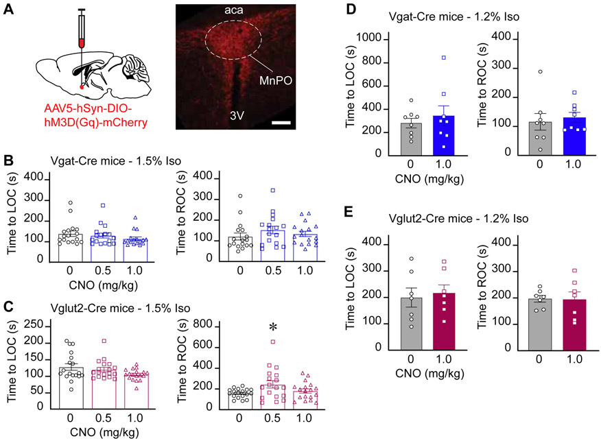
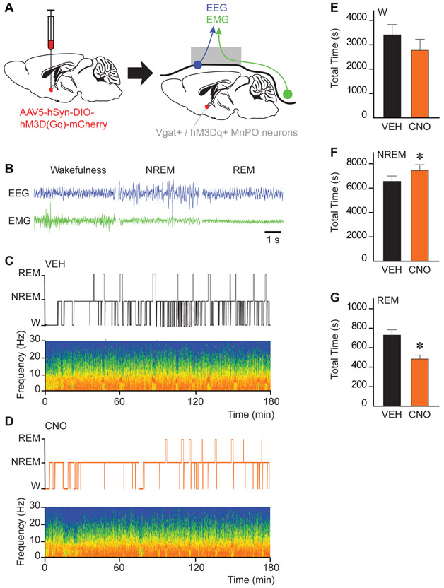
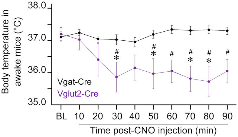
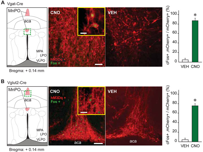
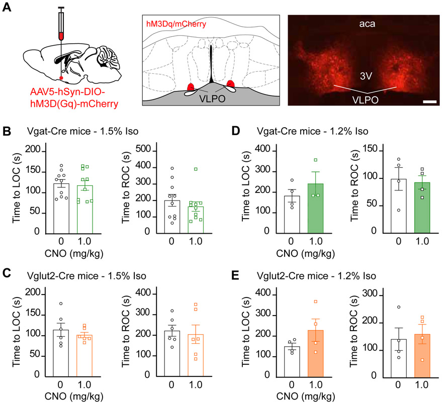
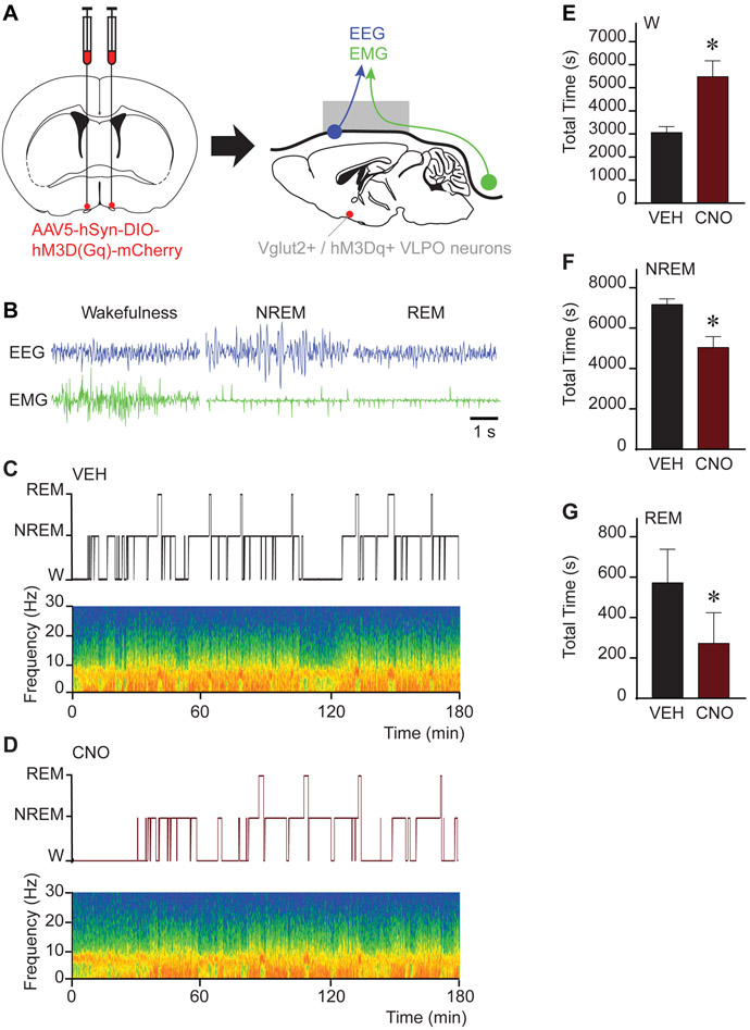
The precise mechanism of general anesthesia remains unclear. In the last two decades, there has been considerable focus on the hypothesis that anesthetics co-opt the neural mechanisms regulating sleep. This hypothesis is supported by ample correlative evidence at the level of sleep-promoting nuclei, but causal investigations of potent inhaled anesthetics have not been conducted. Here, we tested the hypothesis that chemogenetic activation of discrete neuronal subpopulations within the median preoptic nucleus (MnPO) and ventrolateral preoptic nucleus (VLPO) of the hypothalamus would modulate sleep/wake states and alter the time to loss and resumption of consciousness associated with isoflurane, a potent halogenated ether in common clinical use. We show that activating MnPO/VLPO GABAergic or glutamatergic neurons does not alter anesthetic induction or recovery time. However, activation of these neuronal subpopulations did alter sleep-wake architecture. Notably, we report the novel finding that stimulation of VLPO glutamatergic neurons causes a strong increase in wakefulness. We conclude that activation of preoptic GABAergic or glutamatergic neurons that increase sleep or wakefulness does not substantively influence anesthetic state transitions. These data indicate that the correlative evidence for a mechanistic overlap of sleep and anesthesia at the level of an individual nucleus might not necessarily have strong causal significance.

Keywords: DREADD; arousal; consciousness; general anesthesia; isoflurane; sleep; wakefulness.


Conflict of interest statement

Declaration of Interests The authors declare no competing interests.

Figures

Figure 1.
Figure 1.. Chemogenetic stimulation of GABAergic and glutamatergic neurons in the MnPO did not alter anesthetic state transitions
A. Schematic representation of AAV injection into the median preoptic nucleus (MnPO). mCherry (red) indicates the expression of the excitatory designer receptor hM3Dq within the MnPO of a Vglut2-Cre mouse. B. Time to loss and resumption of consciousness (left and right panels, respectively) in Vgat-Cre mice exposed to 1.5% isoflurane. C. Time to loss and resumption of consciousness (left and right panels, respectively) in Vglut2-Cre mice exposed to 1.5% isoflurane. D and E show the time to loss and resumption of consciousness (left and right panels, respectively) in Vgat-Cre and Vglut2-Cre mice exposed to 1.2% isoflurane. The asterisk in C indicates a significant difference (P < 0.05) from vehicle control (0 mg/kg CNO). Statistical comparisons were conducted using a Friedman test followed -when applicable- by a post hoc Dunn’s (B and C) or a two-tailed Wilcoxon test (D and E). Abbreviations: aca, anterior commissure; CNO, clozapine-N-oxide; LOC, loss of consciousness; ROC, resumption of consciousness; 3V, third ventricle. Calibration bar in A, 50 μm. Data in B-E are shown as mean ± standard error of the mean. See also Figures S1, S2 and S4.
Figure 2.
Figure 2.. Chemogenetic stimulation of GABAergic neurons in the MnPO increased NREM sleep and decreased REM sleep
A. Schematic representation of AAV injection into the median preoptic nucleus (MnPO) for expression of the excitatory designer receptor hM3Dq. Following the expression of hM3Dq receptors in the MnPO, mice were implanted with electrodes for recording the electroencephalogram (EEG; blue) and electromyogram (EMG; green). B. Representative EEG and EMG traces recorded from the same Vgat-Cre mouse during wakefulness, non-rapid eye movement (NREM) sleep and rapid eye movement (REM) sleep. C. and D. Hypnograms displaying the time course of sleep and wakefulness during the 3-h recording period. The spectrograms below each hypnogram show state-specific changes in frontal power density (red = maximum, blue = minimum) across the respective recording session. Time 0 on the abscissa indicates the time at which vehicle (VEH) or clozapine-N-oxide (CNO; 1.0 mg/kg) was injected. E, F and G show the effect of CNO administration on the duration (in s) of wakefulness (W), NREM sleep and REM sleep over the 3-h recording period. Asterisks indicate a significant difference (P < 0.05) from vehicle control using a one-tailed Wilcoxon test. Data in E-G are shown as mean ± standard error of the mean. See also Figures S3 and S4
Figure 3.
Figure 3.. Stimulation of glutamatergic (but not GABAergic) neurons in the MnPO produced hypothermia in awake mice
The figure plots the change in core body temperature in awake mice before and after an intraperitoneal injection of clozapine-N-oxide (CNO; 1.0 mg/kg). Asterisks indicate significant differences (P < 0.05) from baseline and hashtag symbols indicate significant differences between mouse lines. Statistical comparisons were conducted using a two-way ANOVA with Bonferroni’s correction for multiple comparisons. The gray area indicates the time period (60 to 90 minutes) during which the time to loss and resumption of consciousness was assessed in anesthesia experiments. Data are shown as mean ± standard error of the mean. See also Figure S4.
Figure 4.
Figure 4.. Verification of MnPO Vgat+ and Vglut2+ cell activation by CNO
A and B. cFos expression (green nuclei) in mCherry expressing (red) neurons within the MnPO of Vgat-Cre and Vglut2-Cre mice, 90 minutes after an intraperitoneal injection of clozapine-N-oxide (CNO; 1.0 mg/kg) or vehicle solution (VEH). The bar graphs plot the percentage (mean + standard error of the mean) of mCherry expressing neurons that also express cFos, over the total number of mCherry positive cells after vehicle or CNO administration. Asterisks indicate a significant difference (P < 0.05) from vehicle control using a one-tailed Mann Whitney test. The strong increase in cFos expression in GABAergic (Vgat+) and glutamatergic (Vglut2+) neurons provides further validation of hM3Dq receptors in the MnPO. Abbreviations: aca, anterior commissure; LPO, lateral preoptic area; MnPO, median preoptic nucleus; MPA, medial preoptic area; VLPO, ventrolateral preoptic nucleus. Calibration bars in A and B, 50 μm and 20 μm (inset).
Figure 5.
Figure 5.. Chemogenetic stimulation of GABAergic and glutamatergic neurons in the VLPO did not alter anesthetic state transitions
A. Schematic representation of bilateral AAV injections into the ventrolateral preoptic nucleus (VLPO). mCherry (red) indicates the expression of the excitatory designer receptor hM3Dq within the VLPO of a Vglut2-Cre mouse. B. Time to loss and resumption of consciousness (left and right panels, respectively) in Vgat-Cre mice exposed to 1.5% isoflurane. C. Time to loss and resumption of consciousness (left and right panels, respectively) in Vglut2-Cre mice exposed to 1.5% isoflurane. D and E show the time to loss and resumption of consciousness (left and right panels, respectively) in Vgat-Cre and Vglut2-Cre mice exposed to 1.2% isoflurane. Statistical comparisons were conducted using two-tailed Wilcoxon test. Abbreviations: aca, anterior commissure; CNO, clozapine-N-oxide; LOC, loss of consciousness; VLPO, ventrolateral preoptic nucleus; ROC, resumption of consciousness; 3V, third ventricle. Calibration bar in A, 50 μm. Data in B-E are shown as mean ± standard error of the mean. See also Figures S2 and S5.
Figure 6.
Figure 6.. Chemogenetic stimulation of glutamatergic neurons in the VLPO increased wakefulness and decreased sleep
A. Schematic representation of bilateral AAV injections into the ventrolateral preoptic nucleus (VLPO) for expression of the excitatory designer receptor hM3Dq. Following the expression of hM3Dq receptors in the VLPO, mice were implanted with electrodes for recording the electroencephalogram (EEG; blue) and electromyogram (EMG; green). B. Representative EEG and EMG signals recorded from the same Vglut2-Cre mouse during wakefulness, non-rapid eye movement (NREM) sleep and rapid eye movement (REM) sleep. C. and D. Hypnograms displaying the time course of sleep and wakefulness during the 3-h recording period. The spectrograms below each hypnogram show state-specific changes in frontal power density (red = maximum, blue = minimum) across the respective recording session. Time 0 on the abscissa indicates the time at which vehicle (VEH) or clozapine-N-oxide (CNO; 1.0 mg/kg) was injected. E, F and G show the effect of CNO administration on the duration (in s) of wakefulness (W), NREM sleep and REM sleep over the 3-h recording period. Asterisks indicate a significant difference (P < 0.05) from vehicle control using a two-tailed Wilcoxon test. Data in E-G are shown as mean ± standard error of the mean. See also Figures S3 and S5.

Comment in

References

    1. Lydic R, and Biebuyck JF (1994). Sleep neurobiology: relevance for mechanistic studies of anaesthesia. Br J Anaesth 72, 506–508. - PubMed
    1. Moore JT, Chen J, Han B, Meng QC, Veasey SC, Beck SG, and Kelz MB (2012). Direct activation of sleep-promoting VLPO neurons by volatile anesthetics contributes to anesthetic hypnosis. Curr Biol 22, 2008–2016. - PMC - PubMed
    1. Nelson LE, Guo TZ, Lu J, Saper CB, Franks NP, and Maze M (2002). The sedative component of anesthesia is mediated by GABA(A) receptors in an endogenous sleep pathway. Nat Neurosci 5, 979–984. - PubMed
    1. Lu J, Nelson LE, Franks N, Maze M, Chamberlin NL, and Saper CB (2008). Role of endogenous sleep-wake and analgesic systems in anesthesia. J Comp Neurol 508, 648–662. - PMC - PubMed
    1. Tung A, Bluhm B, and Mendelson WB (2001). The hypnotic effect of propofol in the medial preoptic area of the rat. Life Sci 69, 855–862. - PubMed

Publication types

LinkOut - more resources