. 2020 Feb 21;15(2):e0224584.
doi: 10.1371/journal.pone.0224584. eCollection 2020.

Evaluation of systemic inflammatory response and lung injury induced by Crotalus durissus cascavella venom

Affiliations

Evaluation of systemic inflammatory response and lung injury induced by Crotalus durissus cascavella venom

Elen Azevedo et al. PLoS One. .

Abstract

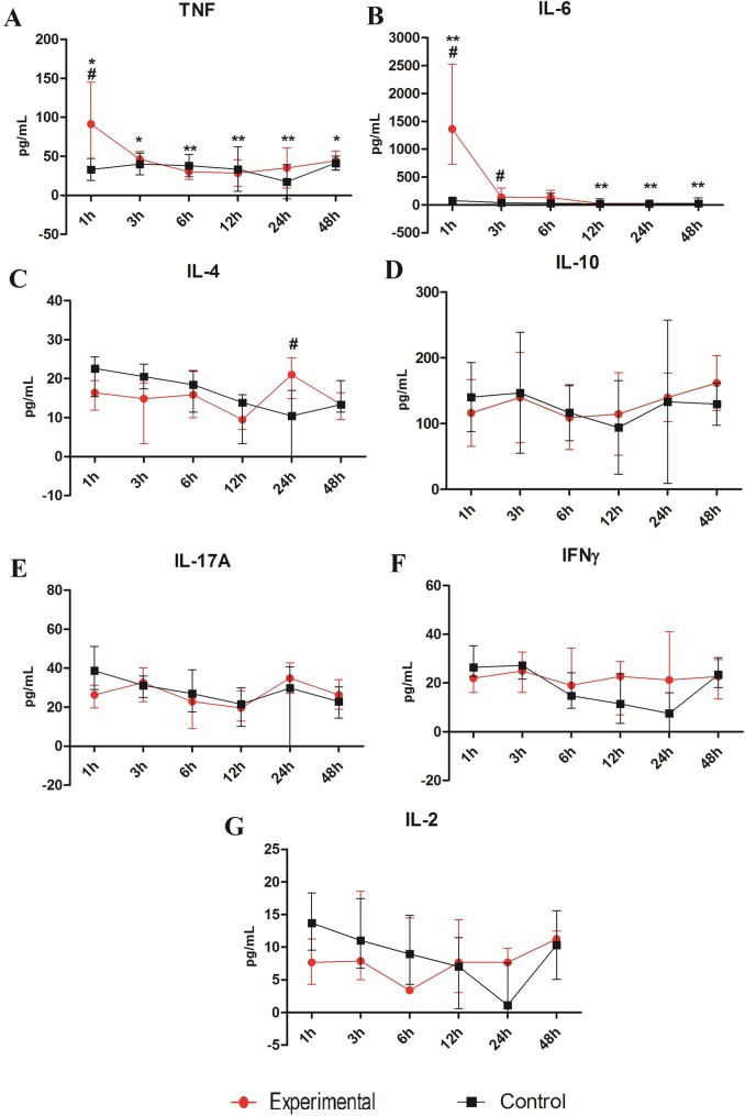
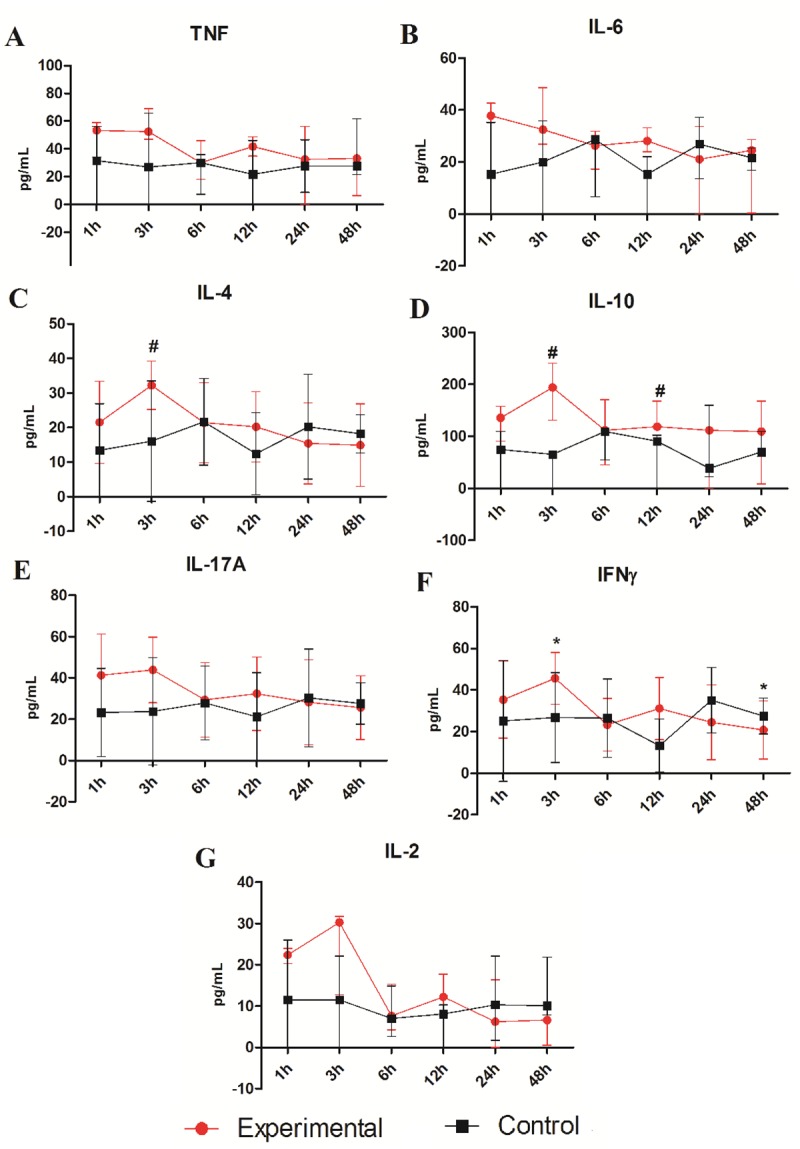
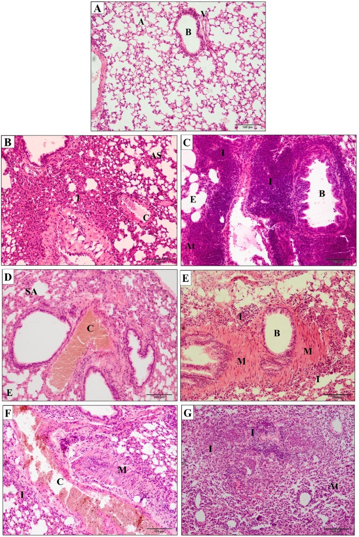
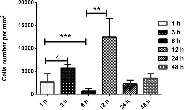
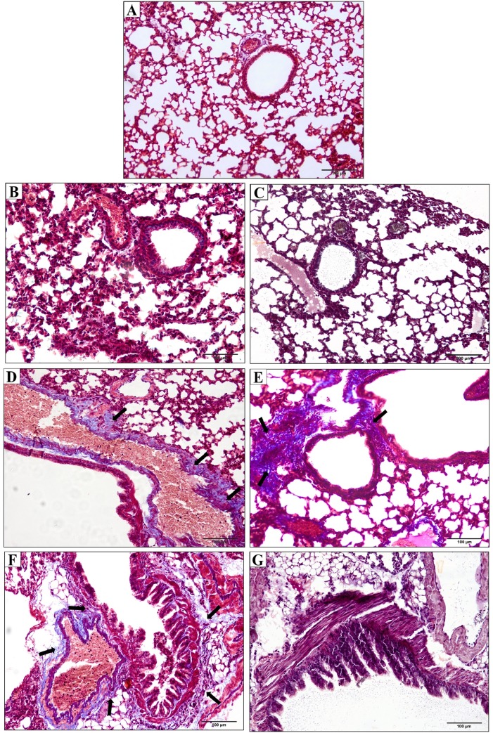
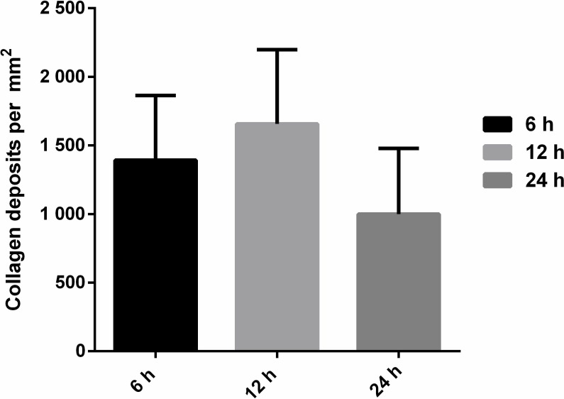
This study investigated the systemic inflammatory response and mechanism of pulmonary lesions induced by Crotalus durissus cascavella venom in murine in the state of Bahia. In order to investigate T helper Th1, Th2 and Th17 lymphocyte profiles, we measured interleukin (IL) -2, IL-4, IL-6, IL-10, IL-17, tumor necrosis factor (TNF) and interferon gamma (IFN-γ) levels in the peritoneal fluid and macerated lungs of mice and histopathological alterations at the specific time windows of 1h, 3h, 6h, 12h, 24h and 48h after inoculation with Crotalus durissus cascavella venom. The data demonstrated an increase of acute-phase cytokines (IL-6 and TNF) in the first hours after inoculation, with a subsequent increase in IL-10 and IL-4, suggesting immune response modulation for the Th2 profile. The histopathological analysis showed significant morphological alterations, compatible with acute pulmonary lesions, with polymorphonuclear leukocyte (PMN) infiltration, intra-alveolar edema, congestion, hemorrhage and atelectasis. These findings advance our understanding of the dynamics of envenomation and contribute to improve clinical management and antiophidic therapy for individuals exposed to venom.


Conflict of interest statement

The authors have declared that no competing interests exist.

Figures

Fig 1
Fig 1. Mice and their clinical manifestations.
Control group was inoculated (i.p.) with 500μL saline solution (A). Experimental group inoculated (i.p.) with 50μg/kg Crotalus durissus cascavella venom diluted in 500μL saline solution (B-D): Initial clinical symptoms observed were pruritus, wound licking (B) and nesting behavior (C); after three hours the animals presented lethargy and respiratory distress (D); 24 hours after inoculation, there were intense abdominal contractions and thoracic effort (E).
Fig 2
Fig 2
Cytokine levels TNF (A), IL-6 (B), IL-10 (C), IL-4 (D), IL-17A (E), IFN-γ (F) and IL-2 (G) measured in peritoneal fluid of the Swiss mice at different time 1, 3, 6, 12, 24 and 48 hours. The mice were inoculated via i.p with 50μg/kg Crotalus durissus cascavella venom diluted in 500μL saline solution. The control group animals were inoculated with 500μL sterile saline solution. Each point represents the mean–SD (A and C) and median—IQR (B, D, E, F and G) of animals per group. *p< 0.05 and **p<0.01 in relation to the venom treatment times and #p<0.05 when compared to the control group.
Fig 3
Fig 3
Cytokine levels TNF (A), IL-6 (B), IL-4 (C), IL-10 (D), IL-17A (E), IFN-γ (F) and IL-2 (G) measured in the macerated lungs of the Swiss mice at different time 1, 3, 6, 12, 24 and 48 hours. The mice were inoculated via i.p with 50μg/kg Crotalus durissus cascavella venom diluted in 500μL saline solution. The control group animals were inoculated with 500μL sterile saline solution. Each point represents the mean–SD (C, E and F) and median–IQR (A, B, D and G) of animals per group. *p< 0.05 in relation to the venom treatment times and #p<0.05 when compared to the control group.
Fig 4
Fig 4. Photomicrographs of pulmonary parenchyma of mice.
(A) Control group inoculated with 500μL sterile saline solution presented preserved pulmonary architecture (A–alveolus; B–bronchial; V–vessels). (B–G) Experimental groups inoculated with 50μg/kg Crotalus durissus cascavella venom at different observation time: (B) 1h; (C) 3h; (D) 6h; (E) 12h; (F) 24h; (G) 48h. (I–Inflammatory infiltrates; AS–alveolar septum thickening; C–vascular congestion; E–emphysema; At–atelectasis; M—bronchial muscle distension).
Fig 5
Fig 5. Morphometric analysis of the frequency of polymorphonuclear cells in inflammatory infiltrates in pulmonary parenchyma of Swiss mice.
Experimental groups inoculated with 50μg/kg Crotalus durissus cascavella venom at different observation time showed significant differences with p = 0,00159 (1 and 3 hours); p = 0,00317 (1 and 6 hours); p = 0,0079 (3, 6, 12 and 24 hours); p = 0,00317 (3 and 48 hours); p = 0,0079 (6 and 12 horas); p = 0,0159 (6 and 24 hours); p = 0,0079 (6 and 48 hours); p = 0,0079 (12 and 24/ 12 and 48 hours). No statistical difference was showed between concentrations at 24 and 48 hours. Statistical analysis was performed by the bilateral Mann—Whitney Test. *p<0,05 were considered statistically significant.
Fig 6
Fig 6. Photomicrographs of pulmonary parenchyma of mice stained with Masson’s trichrome.
(A) Control group inoculated with 500μL sterile saline. (B-G). Experimental groups inoculated with 50μg/kg Crotalus durissus cascavella venom at different observation time: (B) 1h; (C) 3h; (D) 6h; (E) 12h; (F) 24h; (G) 48h. The arrows indicate collagen deposits (stained in blue) in the peribronchial and perivascular regions (Masson’s trichrome staining).
Fig 7
Fig 7. Quantification of collagen deposits in Swiss mice inoculated with 50μg/kg Crotalus durissus cascavella venom at different observation time.
Evaluation of collagen deposits by Masson's trichrome staining revealed no significant differences at any of the correlated points. Statistical analysis performed by the bilateral Mann-Whitney test. p<0,05 was considered statistically significant.

References

    1. World Health Organization. Rabies and envenomings. A neglected public health issue: report of a consultative meeting. [Internet]. World Health Organization, editor. Geneva; 2007. 32 p. Available from: https://apps.who.int/iris/handle/10665/43858
    1. Barravieira B, Lomonte B, Tarkowski A, Hanson LÃ, Meira DA. Acute-phase reactions, including cytokines, in patients bitten by Bothrops and Crotalus snakes in Brazil. J Venom Anim Toxins [Internet]. 1995;1:11–22. Available from: http://www.scielo.br/scielo.php?script=sci_arttext&pid=S0104-79301995000...
    1. Biondi I. Caracterização biológica e bioquímica da peçonha de Crotalus durissus cascavella no Estado da Bahia. State University of Feira de Santana; 2009.
    1. Petricevich VL, Teixeira CFP, Tambourgi D V, Gutiérrez JM. Increments in serum cytokine and nitric oxide levels in mice injected with Bothrops asper and Bothrops jararaca snake venoms. Toxicon [Internet]. 2000;38(9):1253–66. Available from: http://www.sciencedirect.com/science/article/pii/S0041010199002275 10.1016/s0041-0101(99)00227-5 - DOI - PubMed
    1. Luna K, Melo C, Pascoal VPM, Martins-Filho OA, Pereira VRA. Bothrops erythromelas snake venom induces a proinflammatory response in mice splenocytes. Int J Interf Cytokine Mediat Res. 2011. January 1;3:9–18.

Publication types